Matt Tyska (@tyskalabactual) 's Twitter Profile
Matt Tyska

@tyskalabactual

Investigating cytoskeletal control of cell morphology @VanderbiltU using tubes and microscopes. @VUCellImaging @VanderbiltCDB @VUBasicSciences

ID: 1113553373608009728

linkhttps://lab.vanderbilt.edu/tyska-lab/ calendar_today03-04-2019 21:26:27

1,1K Tweet

6,6K Followers

1,1K Following

Mira Krendel (@kidneymyosin) 's Twitter Profile Photo

Postdoctoral position to study myosin functions in immune cells and kidney disease at Upstate Medical University in Syracuse, NY. Affordable living, collaborative environment, and opportunities to use advanced microscopy and protein analysis techniques. bit.ly/4behKQK

Jordan Beach (@myosincity) 's Twitter Profile Photo

The search is on.... We are recruiting at the assistant professor level! Come join us!!! Assistant Professor job with Loyola University of Chicago - Cell and Molecular Physiology Department | 12818067 (nature.com)

Vanderbilt Cell Imaging Shared Resource (@vucellimaging) 's Twitter Profile Photo

We're still obsessed with the #cicadas over here in the CISR. So, here are hair-like projections from a local Vanderbilt cicada leg. Confocal, autofluorescence, 20x, 200 um total, Z projection. #FluorescenceFriday 🔬🪰

We're still obsessed with the #cicadas over here in the CISR. So, here are hair-like projections from a local Vanderbilt cicada leg. Confocal, autofluorescence, 20x, 200 um total, Z projection. #FluorescenceFriday 🔬🪰
Dr. Samantha Stehbens PhD (@samstehbens) 's Twitter Profile Photo

A migrating keratinocyte expressing the microtubule binding protein eGFP-CLASP2 that regulates cell migration. Can you guess when we add the microtubule depolymerising drug? #cytoskeleton #migration

epithelial mechanics fan club (@epimechfc) 's Twitter Profile Photo

Collective cell motion has many forms, but rotation is the coolest of them all. I'm Nimesh Chahare, and in this short🧵, I'll highlight some instances of global tissue rotation like this one from BauschLab

Emma Koory (@emmakoory) 's Twitter Profile Photo

First #FluorescenceFriday as a ✨PhD candidate✨!! This is a cardiomyocyte stained for sarcomere Z-lines (alpha actinin, magenta) and M-lines (titin, yellow). Imaging these guys never gets old!

First #FluorescenceFriday as a ✨PhD candidate✨!! This is a cardiomyocyte stained for sarcomere Z-lines (alpha actinin, magenta) and M-lines (titin, yellow). Imaging these guys never gets old!
Matt Tyska (@tyskalabactual) 's Twitter Profile Photo

📢 Heads up local microscopy peeps: Today is the deadline for the CISR “Life is Beautiful” contest 🤩 Send in your good stuff to win STACKS of cash! 💰💰👇👇

Matt Tyska (@tyskalabactual) 's Twitter Profile Photo

Music city delivered on the fireworks this year 🤩💥💥💥💥 #NashvilleJuly4 #Nashville #fireworks #Tennessee #IndependenceDay #4thofJuly

Weiner Lab (@oweinerlab) 's Twitter Profile Photo

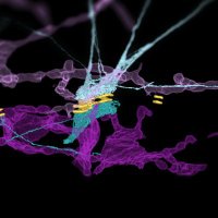
✨Pre-print alert!! ✨ Check out Muziyue’s incredible thesis work ➡️ “The WAVE complex forms linear arrays at negative membrane curvature to instruct lamellipodia formation”

MohanBalasubramanian (@sporemohan) 's Twitter Profile Photo

Please feel free to retweet I have a post-doctoral position to measure cytokinetic forces in collaboration with Justin Molloy in Warwick. warwick-careers.tal.net/vx/lang-en-GB/…

Kowalczyk Lab (@celljxn) 's Twitter Profile Photo

This was a wonderful project to work on with Alexa Mattheyses and Navaneetha Bharathan, PhD. Nav Bharathan led this effort to review the history of desmosome imaging, from the mid 1800s to today. It is inspiring to review the work of early imaging scientists and pioneers of EM.
Alushin_Lab (@alushin_lab) 's Twitter Profile Photo

Particularly excited to share our latest work, where we visualize how myosin motor forces alter the structure of actin. We discovered a spiral form of the filament which can be recognized by force-sensitive actin binding proteins. biorxiv.org/content/10.110…