NEMI (@projectnemi) 's Twitter Profile
NEMI

@projectnemi

NEMI (Netherlands Electron Microscopy Infrastructure )

ID: 1159397131050004481

linkhttp://nemi.microscopie.nl calendar_today08-08-2019 09:33:11

309 Tweet

338 Followers

245 Following

NEMI (@projectnemi) 's Twitter Profile Photo

🍯"Nano-honeycomb"🐝by Egemen Deniz Eren, TU Eindhoven @tueindhoven Intricate #nanostructures of raspberry supraparticles, forming a multiscale hierarchy reminiscent of a honeycomb!🍯 🔬 FEI Quanta 600 FEG (#SEM), TU Eindhoven @nemiproject Thermo Scientific #Microscopy

🍯"Nano-honeycomb"🐝by Egemen Deniz Eren, TU Eindhoven @tueindhoven 

Intricate #nanostructures of raspberry supraparticles, forming a multiscale hierarchy reminiscent of a honeycomb!🍯

🔬 FEI Quanta 600 FEG (#SEM), TU Eindhoven

@nemiproject <a href="/thermosci/">Thermo Scientific</a> #Microscopy
NEMI (@projectnemi) 's Twitter Profile Photo

🌌 "Beyond Darkness" 🌟 by Alessandro Borsellini, Leiden UMC LUMC Leiden 🧬Delve into the intricate world of #DNA repair with this high-resolution structure of the MutS protein. 🔬Titan Krios (Microscopy & Spectroscopy) at Netherlands Centre for Electron Nanoscopy (NeCEN), NeCEN
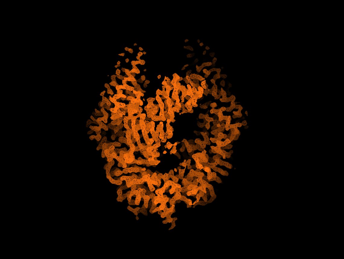
🌌 "Beyond Darkness" 🌟 by Alessandro Borsellini, Leiden UMC <a href="/LUMC_Leiden/">LUMC Leiden</a>

🧬Delve into the intricate world of #DNA repair with this high-resolution structure of the MutS protein.

🔬Titan Krios (<a href="/thermosciEMSpec/">Microscopy & Spectroscopy</a>) at Netherlands Centre for Electron Nanoscopy (NeCEN), <a href="/NeCEN_2p0/">NeCEN</a>
NEMI (@projectnemi) 's Twitter Profile Photo

"A Collage of Correlation" 🖼️by Jan van der Beek, UMC Utrecht The green + red fluorescent signals of Rab5 + Rab7 overlay the complex ultrastructure of #endosomes + #lysosomes, connecting their molecular + structural identity. 🔬Tecnai T12 (#EM) & Deltavision (light #microscopy)

"A Collage of Correlation" 🖼️by Jan van der Beek, <a href="/UMCUtrecht/">UMC Utrecht</a>

The green + red fluorescent signals of Rab5 + Rab7 overlay the complex ultrastructure of #endosomes + #lysosomes, connecting their molecular + structural identity.

🔬Tecnai T12 (#EM) &amp; Deltavision (light #microscopy)
NEMI (@projectnemi) 's Twitter Profile Photo

"The beauty of forbidden symmetry in binary supraparticles" by Da Wang, Utrecht University (@utrechtuniversity) 🟢🔵A mixture of 2 types of quantum dots self-assembled into binary icosahedral supraparticles from spherical confinement. 🔬Talos F200 in #STEM mode Microscopy & Spectroscopy
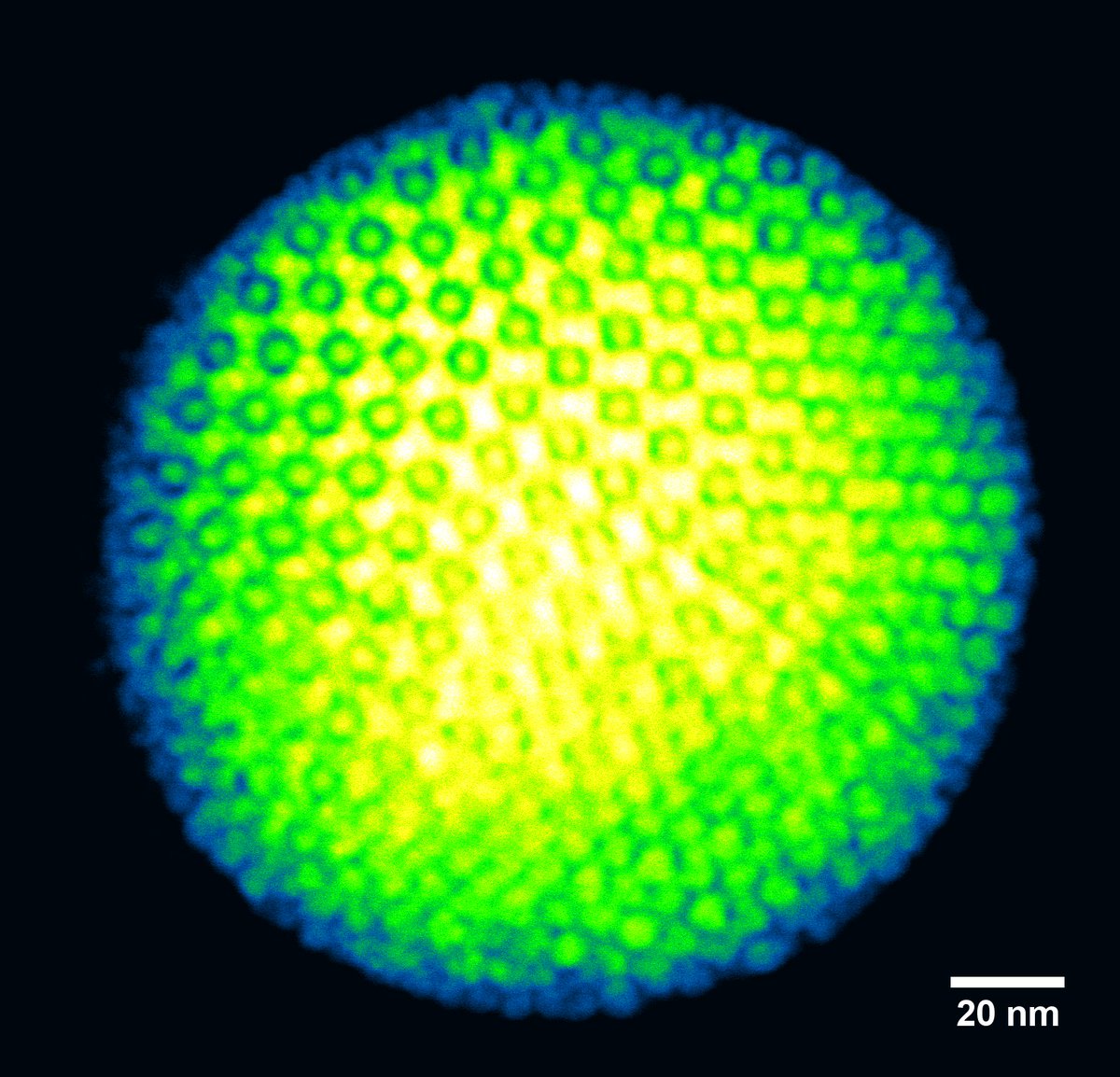
"The beauty of forbidden symmetry in binary supraparticles" by Da Wang, Utrecht University (@utrechtuniversity)

🟢🔵A mixture of 2 types of quantum dots self-assembled into binary icosahedral supraparticles from spherical confinement.

🔬Talos F200 in #STEM mode <a href="/thermosciEMSpec/">Microscopy & Spectroscopy</a>
NEMI (@projectnemi) 's Twitter Profile Photo

📆 May image of the month "Roses" by Pekka Kujala, UMC Utrecht (UMC Utrecht). 🌹Explore rosette-like, swirled membranes of late endosomes (red) in Drosophila midgut. 🔬 Imaged with a JEOL 1010 #TEM (JEOL EUROPE) at the Cell Microscopy Core, UMC Utrecht (UMC Utrecht)
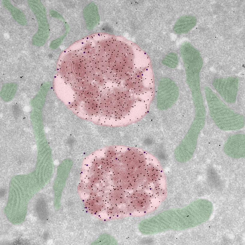
📆 May image of the month "Roses" by Pekka Kujala, UMC Utrecht (<a href="/UMCUtrecht/">UMC Utrecht</a>).

🌹Explore rosette-like, swirled membranes of late endosomes (red) in Drosophila midgut. 

🔬 Imaged with a JEOL 1010 #TEM (<a href="/JEOLEUROPE/">JEOL EUROPE</a>) at the Cell Microscopy Core, UMC Utrecht (<a href="/UMCUtrecht/">UMC Utrecht</a>)
NEMI (@projectnemi) 's Twitter Profile Photo

✨✈️NEMI Travel Grant 🚊✨ NEMI is offering financial support for Early Career Researchers traveling to conferences/workshops! NEMI will cover 75% of the expenses (conference/workshop fee, travel, & housing). 🔗Link to the form via our website: nemi.microscopie.nl/news/nemi-trav…

✨✈️NEMI Travel Grant 🚊✨ <a href="/ProjectNemi/">NEMI</a> is offering financial support for Early Career Researchers  traveling to conferences/workshops! NEMI will cover 75% of the expenses (conference/workshop fee, travel, &amp; housing).

🔗Link to the form via our website: nemi.microscopie.nl/news/nemi-trav…
NEMI (@projectnemi) 's Twitter Profile Photo

"Spying on the Secrets of Sperm"- Miguel Ricardo Leung, Marc Roelofs, Tzviya Zeev-Ben-Mordehai, Utrecht University Explore the unique structures of sperm centrioles with a computational slice through a cryo-tomogram of a boar sperm cell. 🔬Titan Krios, Microscopy & Spectroscopy at NeCEN

"Spying on the Secrets of Sperm"- Miguel Ricardo Leung, Marc Roelofs, Tzviya Zeev-Ben-Mordehai, <a href="/UniUtrecht/">Utrecht University</a> 

Explore the unique structures of sperm centrioles with a computational slice through a cryo-tomogram of a boar sperm cell. 

🔬Titan Krios, <a href="/thermosciEMSpec/">Microscopy & Spectroscopy</a> at <a href="/NeCEN_2p0/">NeCEN</a>
NEMI (@projectnemi) 's Twitter Profile Photo

🔍 "Unveiling Oxygen Deficiency" by Majid Ahmadi, University of Groningen. 🌟 Witness the formation of oxygen-deficient #perovskite in LSMO film during an in-situ biasing experiment through an iDPC #STEM image. 🔬 Imaged at University of Groningen, (University of Groningen)
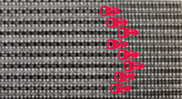
🔍 "Unveiling Oxygen Deficiency" by Majid Ahmadi, University of Groningen.

🌟 Witness the formation of oxygen-deficient #perovskite in LSMO film during an in-situ biasing experiment through an iDPC #STEM image.

🔬 Imaged at University of Groningen, (<a href="/univgroningen/">University of Groningen</a>)
NEMI (@projectnemi) 's Twitter Profile Photo

🌐"HeLa Cells Exposed to Cisplatin" by Nicole Pirozzi, UMCG Discover the intricate interactions of cisplatin within HeLa cells in culture.🔬This Talos F200x #EDX overlay reveals osmium (r), platinum (g), & phosphorus (b), highlighting sequestered platinum in a terminal lysosome

🌐"HeLa Cells Exposed to Cisplatin" by Nicole Pirozzi, <a href="/umcg/">UMCG</a>
Discover the intricate interactions of cisplatin within HeLa cells in culture.🔬This Talos F200x #EDX overlay reveals osmium (r), platinum (g), &amp; phosphorus (b), highlighting sequestered platinum in a terminal lysosome
NEMI (@projectnemi) 's Twitter Profile Photo

🎈"Micro-balloon on a Nail Bed"- Simon Houben & Roel van Raak, TU Eindhoven 🔬 A micro-sized hair array made from a liquid crystalline polymer, supporting a glass microbead. Captured using a FEI 3D Quanta FEG at the Laboratory of Stimuli-responsive Functional Materials & Devices
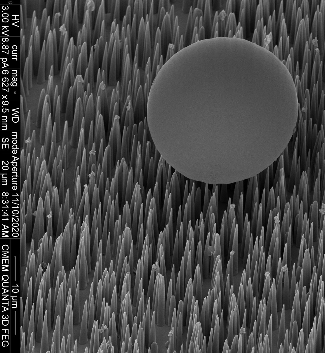
🎈"Micro-balloon on a Nail Bed"- Simon Houben &amp; Roel van Raak, <a href="/TUeindhoven/">TU Eindhoven</a>

🔬 A micro-sized hair array made from a liquid crystalline polymer, supporting a glass microbead. Captured using a FEI 3D Quanta FEG at the Laboratory of Stimuli-responsive Functional Materials &amp; Devices
NEMI (@projectnemi) 's Twitter Profile Photo

🌳"Polls of a Willow Tree"🍃by Ingeborg Schreur, TU Eindhoven 🌿A closer look at the detailed structures of the willow tree's pollen, imaged with a #SEM QUANTA (FEI) at 3 kV. #SEM #WillowTree #Botany #TUEindhoven #Nemi #NemiProject #Microscopy #ElectronMicroscopy
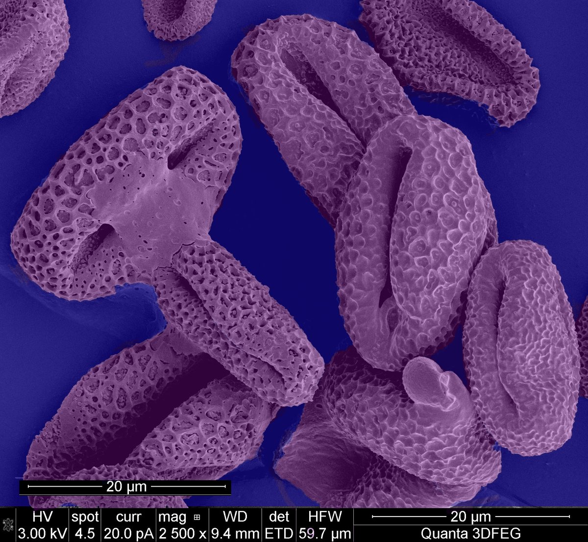
🌳"Polls of a Willow Tree"🍃by Ingeborg Schreur, <a href="/TUeindhoven/">TU Eindhoven</a>

🌿A closer look at the detailed structures of the willow tree's pollen, imaged with a #SEM QUANTA (FEI) at 3 kV.

#SEM #WillowTree #Botany #TUEindhoven #Nemi #NemiProject #Microscopy #ElectronMicroscopy
NEMI (@projectnemi) 's Twitter Profile Photo

🌋 "Volcano Dome" 🌋 by Aya Mahgoub, TU Delft (TU Delft) 🔬 A nanometer-scale volcano dome crafted using focused electron beam induced deposition (#FEBID) serves as the exhaust part of a miniature gas chip, a novel source for ion beam #microscopy. #Nanotechnology #Nemi #TUDelft

🌋 "Volcano Dome" 🌋 by Aya Mahgoub, TU Delft (<a href="/tudelft/">TU Delft</a>)

🔬 A nanometer-scale volcano dome crafted using focused electron beam induced deposition (#FEBID) serves as the exhaust part of a miniature gas chip, a novel source for ion beam #microscopy.

#Nanotechnology #Nemi #TUDelft
NEMI (@projectnemi) 's Twitter Profile Photo

📆Image of the month: June - "World Without Walls" by Joost Willemse, Leiden University 🦠A glimpse into the emergence of L-form bacteria in an E.coli biofilm 🔬 Jeol 7600 #SEM (JEOL EUROPE), Institute of Biology Leiden (IBL) Microscopy unit, Leiden University (Leiden University)

📆Image of the month: June - "World Without Walls" by Joost Willemse, Leiden University 

🦠A glimpse into the emergence of L-form bacteria in an E.coli biofilm

🔬 Jeol 7600 #SEM (<a href="/JEOLEUROPE/">JEOL EUROPE</a>), Institute of Biology Leiden (IBL) Microscopy unit, Leiden University (<a href="/UniLeidenNews/">Leiden University</a>)
NEMI (@projectnemi) 's Twitter Profile Photo

✨✈️NEMI Travel Grant 🚊✨ NEMI is offering financial support for Early Career Researchers traveling to conferences/workshops! #NEMI will cover 75% of the expenses (conference/workshop fee, travel, & housing). 🔗Link to the form via our website: nemi.microscopie.nl/news/nemi-trav…

✨✈️NEMI Travel Grant 🚊✨ <a href="/ProjectNemi/">NEMI</a> is offering financial support for Early Career Researchers  traveling to conferences/workshops! #NEMI will cover 75% of the expenses (conference/workshop fee, travel, &amp; housing).

🔗Link to the form via our website: nemi.microscopie.nl/news/nemi-trav…
NEMI (@projectnemi) 's Twitter Profile Photo

📆Image of the month: July "Caught in a Web" by Cody van der Spek, Amsterdam UMC 🕸️Red blood cells trapped behind a network of fibrin in a human carotid artery. 🔬 Imaged at 3.00 kV with a ZEISS Gemini Sigma 300 #SEM (ZEISS Microscopy), Electron Microscopy Center Amsterdam (EMCA)

📆Image of the month: July "Caught in a Web" by Cody van der Spek, <a href="/amsterdamumc/">Amsterdam UMC</a>

🕸️Red blood cells trapped behind a network of fibrin in a human carotid artery.
 
🔬 Imaged at 3.00 kV with a ZEISS Gemini Sigma 300 #SEM (<a href="/zeiss_micro/">ZEISS Microscopy</a>), Electron Microscopy Center Amsterdam (EMCA)
NEMI (@projectnemi) 's Twitter Profile Photo

✨✈️NEMI Travel Grant 🚊✨ NEMI is offering financial support for Early Career Researchers traveling to conferences/workshops! NEMI will cover 75% of the expenses (conference/workshop fee, travel, & housing). 🔗Link to the form via our website: nemi.microscopie.nl/news/nemi-trav…

✨✈️NEMI Travel Grant 🚊✨ <a href="/ProjectNemi/">NEMI</a> is offering financial support for Early Career Researchers  traveling to conferences/workshops! NEMI will cover 75% of the expenses (conference/workshop fee, travel, &amp; housing).
🔗Link to the form via our website: nemi.microscopie.nl/news/nemi-trav…
NEMI (@projectnemi) 's Twitter Profile Photo

📆Image of the month: August "Floral fireworks" by Luuk M. Moone & Inge van de Ven, TU Eindhoven 🌺Flower-like features revealed after wetting a phenol-urea-formaldehyde coating surface with water and subsequent drying under ambient conditions🌼 🔬Quanta 200 3D-FEG #SEM, CMEM
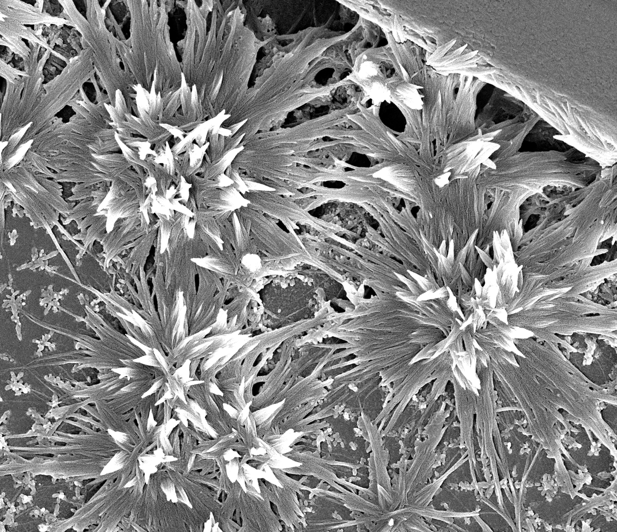
📆Image of the month: August "Floral fireworks" by Luuk M. Moone &amp; Inge van de Ven, <a href="/TUeindhoven/">TU Eindhoven</a>

🌺Flower-like features revealed after wetting a phenol-urea-formaldehyde coating surface with water and subsequent drying under ambient conditions🌼

🔬Quanta 200 3D-FEG #SEM, CMEM
NEMI (@projectnemi) 's Twitter Profile Photo

✨✈️NEMI Travel Grant 🚊✨ NEMI is offering financial support for Early Career Researchers traveling to conferences/workshops! #NEMI will cover 75% of the expenses (conference/workshop fee, travel, & housing). 🔗Link to the form via our website: nemi.microscopie.nl/news/nemi-trav…

✨✈️NEMI Travel Grant 🚊✨ <a href="/ProjectNemi/">NEMI</a> is offering financial support for Early Career Researchers  traveling to conferences/workshops! #NEMI will cover 75% of the expenses (conference/workshop fee, travel, &amp; housing).

🔗Link to the form via our website: nemi.microscopie.nl/news/nemi-trav…
NEMI (@projectnemi) 's Twitter Profile Photo

📆Image of the Month: September "A.I. Colored Slice of Fish" by Roman Koning, LUMC Leiden 🎨Supervised machine learning assisted color segmentation of tissues + organelles of a TEM image of a 5-dpf Zebrafish embryo🐠 🔬Tecnai 12 #TEM, EM Facility, LUMC Leiden #Nemi #biology
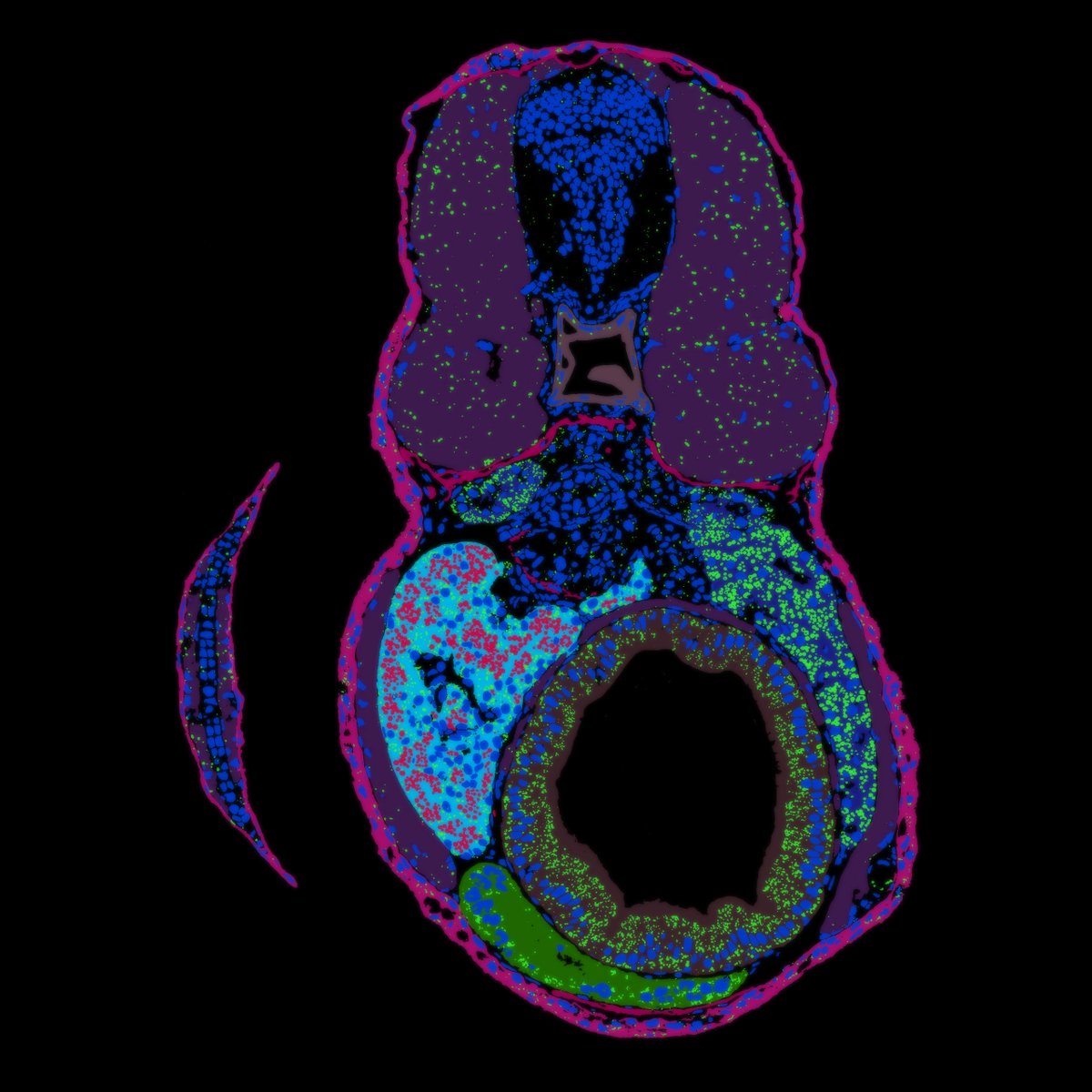
📆Image of the Month: September "A.I. Colored Slice of Fish" by Roman Koning, <a href="/LUMC_Leiden/">LUMC Leiden</a>

🎨Supervised machine learning assisted color segmentation of tissues + organelles of a TEM image of a 5-dpf Zebrafish embryo🐠

🔬Tecnai 12 #TEM, EM Facility, <a href="/LUMC_Leiden/">LUMC Leiden</a>

#Nemi #biology
NEMI (@projectnemi) 's Twitter Profile Photo

✨✈️NEMI Travel Grant 🚊✨ NEMI is offering financial support for Early Career Researchers traveling to conferences/workshops! NEMI will cover 75% of the expenses (conference/workshop fee, travel, & housing). 🔗Link to the form via our website: nemi.microscopie.nl/news/nemi-trav…

✨✈️NEMI Travel Grant 🚊✨ <a href="/ProjectNemi/">NEMI</a> is offering financial support for Early Career Researchers  traveling to conferences/workshops! NEMI will cover 75% of the expenses (conference/workshop fee, travel, &amp; housing).
🔗Link to the form via our website: nemi.microscopie.nl/news/nemi-trav…