Kevin Terretaz (@kwolbachia) 's Twitter Profile
Kevin Terretaz

@kwolbachia

Cells, microscopy, Wolbachia and ImageJ 🔬

ID: 985836950719475713

linkhttps://github.com/kwolbachia calendar_today16-04-2018 11:06:59

1,1K Tweet

759 Takipçi

513 Takip Edilen

BIC (@bic_bordeaux) 's Twitter Profile Photo

#ImageOfTheMonth This image was acquired by J.Angibaud Interdisciplinary Institute for Neuroscience 🧠. Neurons and astrocytes are cultured on a soft polyacrylamide hydrogel. Neurons are labeled for MAP-2 protein (blue), astrocytes for GFAP protein (red) and transfected neurons show GFP expression (green).
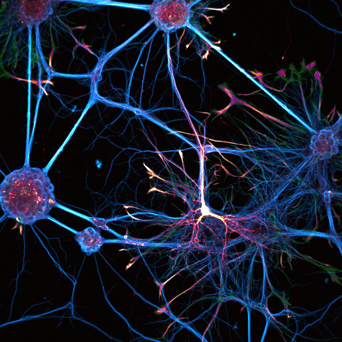
#ImageOfTheMonth
This image was acquired by J.Angibaud <a href="/iins_bordeaux/">Interdisciplinary Institute for Neuroscience 🧠</a>. Neurons and astrocytes are cultured on a soft polyacrylamide hydrogel. Neurons are labeled for MAP-2 protein (blue), astrocytes for GFAP protein (red) and transfected neurons show GFP expression (green).
Abbelight (@abbelight) 's Twitter Profile Photo

Introducing the new 𝗦𝗔𝗙𝗲 platform with an expanded 230x230 µm² FOV, enabling you to visualize larger samples and gather more data in a single acquisition! Ready to explore the largest FOV on the market? Join us at #SFN2024 to see it in action! #Bioimaging #Microscopy

Introducing the new 𝗦𝗔𝗙𝗲 platform with an expanded 230x230 µm² FOV, enabling you to visualize larger samples and gather more data in a single acquisition!

Ready to explore the largest FOV on the market? Join us at #SFN2024 to see it in action! 

#Bioimaging #Microscopy
Stephanie Huang (@stephjhuang) 's Twitter Profile Photo

A Purkinje cell from the Cerebellum labelled with DiI. #FluorescenceFriday I accidentally lopped off the cell body while remounting it, but the branching still looks cool! The image is depicted with Kevin Terretaz's KTZ_bw_Incendio LUT via Christophe Leterrier's NeuroCyto LUTs from ImageJ.

A Purkinje cell from the Cerebellum labelled with DiI. #FluorescenceFriday

I accidentally lopped off the cell body while remounting it, but the branching still looks cool!

The image is depicted with <a href="/kWolbachia/">Kevin Terretaz</a>'s KTZ_bw_Incendio LUT via <a href="/christlet/">Christophe Leterrier</a>'s NeuroCyto LUTs from ImageJ.
FocalPlane (@focalplane_jcs) 's Twitter Profile Photo

On #FluorescenceFriday we’re highlighting our featured image post with Ciarán Butler-Hallissey Ciarán Butler-Hallissey. Check out our post to find out more about the image and learn about Ciarán’s research: focalplane.biologists.com/2024/09/13/fea… #neurons #UExM #Microscopy

On #FluorescenceFriday we’re highlighting our featured image post with Ciarán Butler-Hallissey <a href="/ButlerHallissey/">Ciarán Butler-Hallissey</a>. Check out our post to find out more about the image and learn about Ciarán’s research: focalplane.biologists.com/2024/09/13/fea…
#neurons #UExM #Microscopy
Jason Kirk🔬📷 (@jasonmileskirk) 's Twitter Profile Photo

It's been a hot minute since I posted for #FluorescenceFriday but I finally had a chance to take some inaugural images on our mesoSPIM: Clear Images. Big samples. Open source. and that microscope is FABULOUS! Here is a mouse spinal cord cleared in EZClear showing PRG4 expressing cells. Scale bar is 1mm!!

It's been a hot minute since I posted for #FluorescenceFriday but I finally had a chance to take some inaugural images on our <a href="/MesoSpim/">mesoSPIM: Clear Images. Big samples. Open source.</a> and that microscope is FABULOUS!  Here is a mouse spinal cord cleared in EZClear showing PRG4 expressing cells.  Scale bar is 1mm!!
TJ Krzystek (@lakeerieneuro) 's Twitter Profile Photo

These iPSC-derived motor neuron kinetics really flaunt the dynamic neurite remodeling and outgrowth that occurs in culture🔬 Kinetics taken on a Cytation10 imager US BioTek Laboratories Agilent Technologies Agilent Life Science Inverted Teal LUT by Kevin Terretaz (simply the best!)

Stanislav Vosolsobě (@svosolsobe) 's Twitter Profile Photo

Only 20 minutes is needed for anther opening in #Arabidopsis! Timelapse done by a vertical spinning-disk microscopy on flowers attached to plants. Specific desiccation of anther tissue vs. a fully watered filaments is clearly visible.