
Daniel Frisch, MD
@FrischMd
EP in Philadelphia
ID:1036686322738647041
http://www.jeffersonhealth.org 03-09-2018 18:44:14
90 Tweets
486 Followers
77 Following

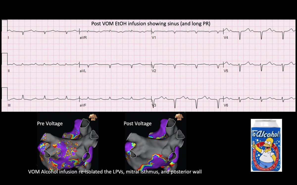
We hoped this would happen but did not expect it...termination of left atrial flutter during VOM ethanol infusion. Hopefully it helps this patient. Nice maps Ryan Coleman maddyferraro Andrew Campadonico Miguel Valderrábano Jefferson Cardiology Fellows


Never a dull moment rounding on EP with Dr. Daniel Frisch, MD. Great teaching with some comedy mixed in. What more could you ask for ???

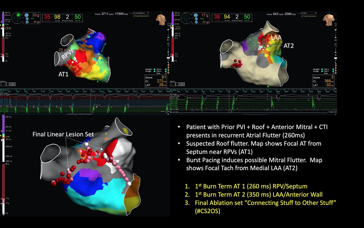
Unusual redo because the previous ablation was intact (PVI, roof, anterior mitral line) yet 2 distinct ATs came from areas near the previous ablation🤔. Termination (x2) was quick, but we decided on linear ablation #CS2OS . Thanks Ryan Coleman Jefferson Cardiology Fellows Abbott Cardiovascular

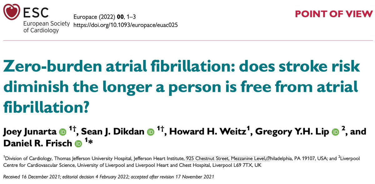
In our editorial, we argue that stroke risk might diminish after a period of AF-free time. Thus, there is value to investigating the time since the last AF event to the future risk of stroke. Daniel Frisch, MD Sean Dikdan Howard Weitz Liverpool Centre for Cardiovascular Science Jefferson Cardiology Fellows
pubmed.ncbi.nlm.nih.gov/35244689/


Great job team. Congratulations!
doi.org/10.1007/s00380… Jefferson Cardiology Fellows Andrew Campadonico Michael Co Joey Junarta Ramya Bodempudi MD

In this interesting case of mitral flutter we were surprised by how proximal the VOM was in the CS with venography. Luckily we had a guide! Thanks Miguel Valderrábano Michael Co Abbott Cardiovascular Jefferson Cardiology Fellows sciencedirect.com/science/articl…
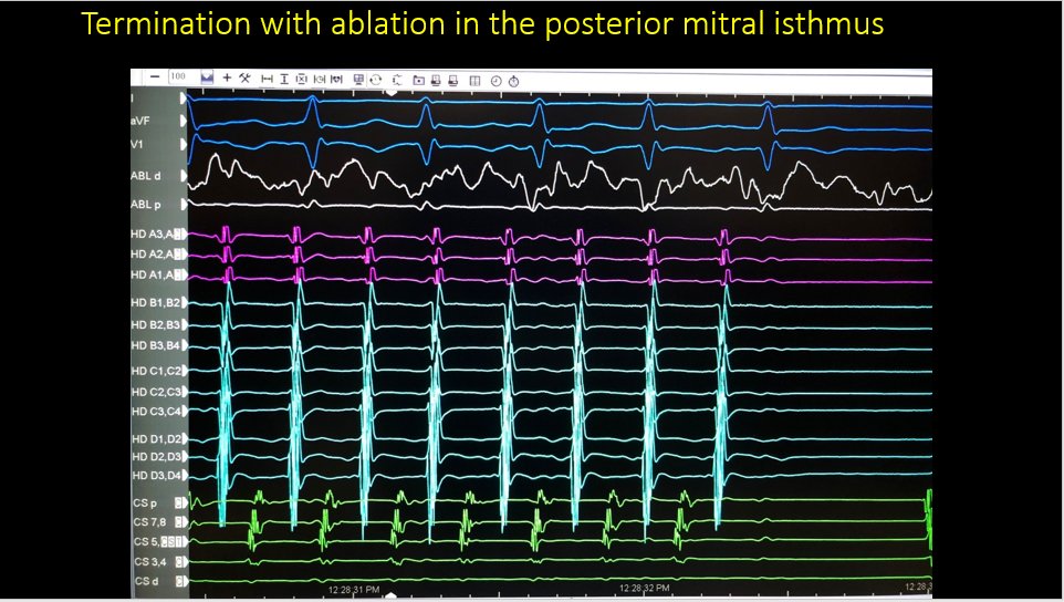

1/
⚡️Want to up your EP game?⚡️
Here is a case-based tweetorial teaching a simple trick to make you look like a pro. #ACCFIT #EPeeps #Cardiotwitter
By Jefferson Cardiology Fellows Noah Haroian, MD PharmD Alexander Hajduczok, MD 🇺🇦🙏 + EP attending Daniel Frisch, MD
Cardiac surgery consult - post op, stable, and this #ECG
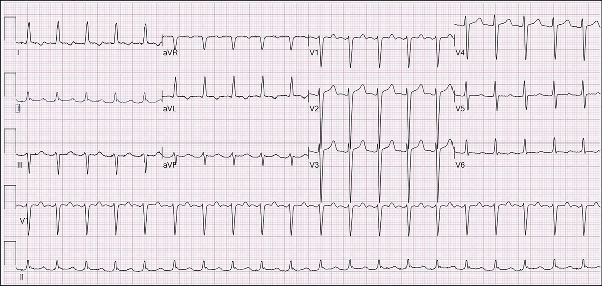

Nice work team! ajconline.org/article/S0002-… AliveCor David E. Albert, M.D Jefferson Cardiology Fellows

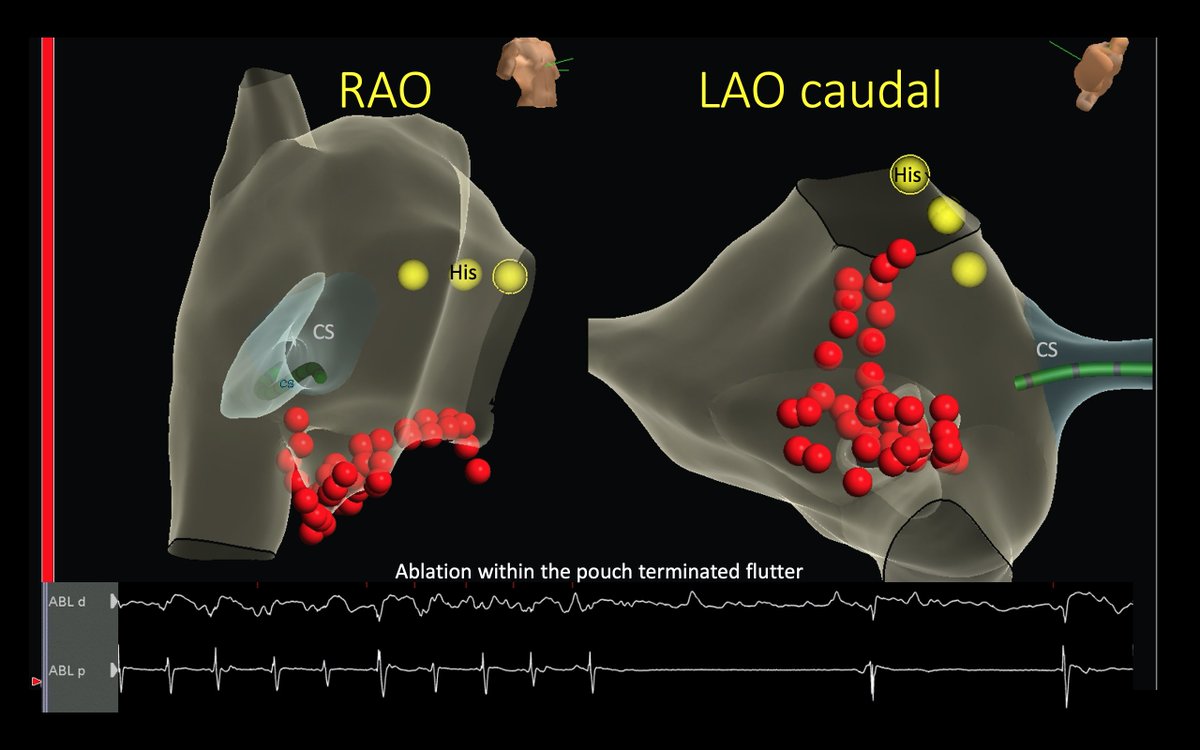
I wonder if ablation here (is safe and) would work for a refractory roof line/flutter Abbott Cardiovascular maddyferraro
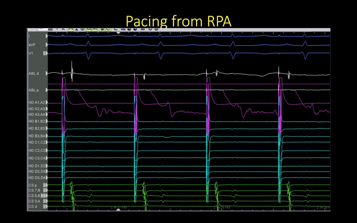

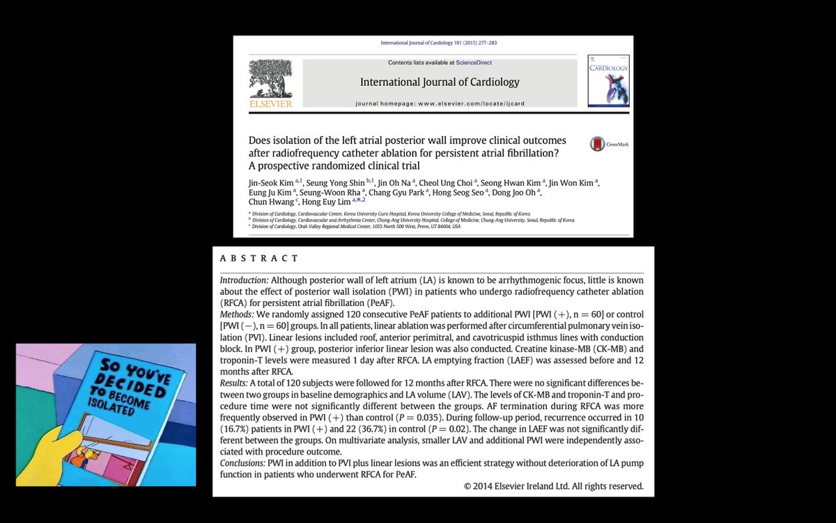
This patient had PAF after prior abl for Persistent AF. PVI was intact. We targeted the posterior wall (PW). An interesting EGM was seen on the grid as the remaining connection and (unusually) we were able to show exit block from the PW Ryan Coleman Abbott Cardiovascular

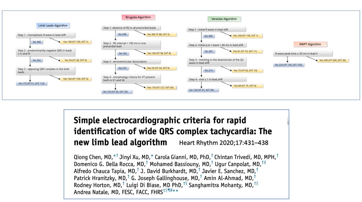
This was an unusual opportunity to record (slow) VT using a Kardia 6L and show the corresponding ECGs. Choose your algorithm… David E. Albert, M.D JMC Jefferson Cardiology Fellows

ICE was essential in recognizing the variant anatomy in this patient. Subsequently, we appreciated it in the other imaging modalities. Thanks Ryan Coleman Abbott Cardiovascular

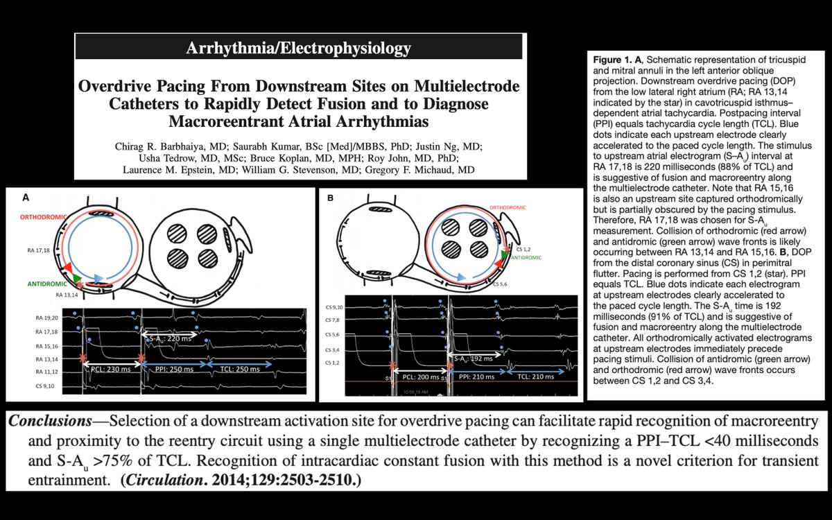
This pt presented for incessant atypical AFL after prior PVI, roof, mitral, and CTI abl. The entrainment observation was helpful in determining reentry as the mechanism. Thanks for the high-density map and high-level insights! Gregory Michaud Abbott Cardiovascular maddyferraro

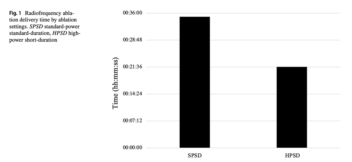
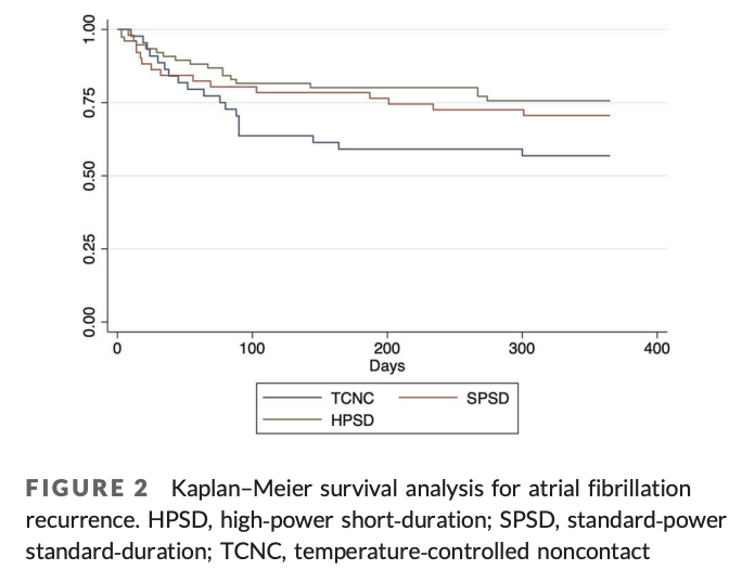
Strong work team! Nice job reviewing our experience with high power short duration ablation for AF (dx.doi.org/10.1111/jce.14…) @Med_Lit_Review Jefferson Cardiology Fellows Andrew Campadonico Abbott Cardiovascular

Congratulations to all the fellows who passed their general cardiology boards. Welcome to the club! Jefferson Cardiology Fellows

Given how anatomically close the RSPV and SVC are, it's no surprise that far-field SVC signals can be recorded from the RSPV. Yet it still can be confusing (to me) Ryan Coleman Abbott Cardiovascular
