Malak Itani
@ItaniMalak
Abdominal Radiologist at Mallinckrodt Institute of Radiology St. Louis - Interested in medical education and healthcare awareness
ID:585943456
20-05-2012 19:48:03
1,9K Tweets
2,1K Followers
1,3K Following
Follow People


Strong presence for Mallinckrodt Institute of Radiology at #SAR24 with 10 speakers from our Abdominal Imaging Section. Don't miss superb talks by Dr. Shetty Anup Shetty our amazing PD, Dr. Mellnick our fearless chief, Drs. Ludwig, Fraum, Ballard, Tsai, Rajput, Hoegger, Lanier, and myself.


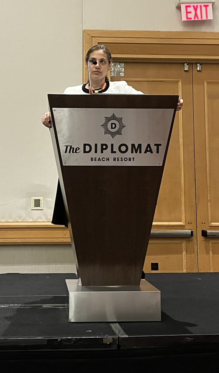
#SAR24 Immunotherapy complications review by the brilliant Malak Itani and Mark Anderson, MD happening now in Atlantic Ballroom 1

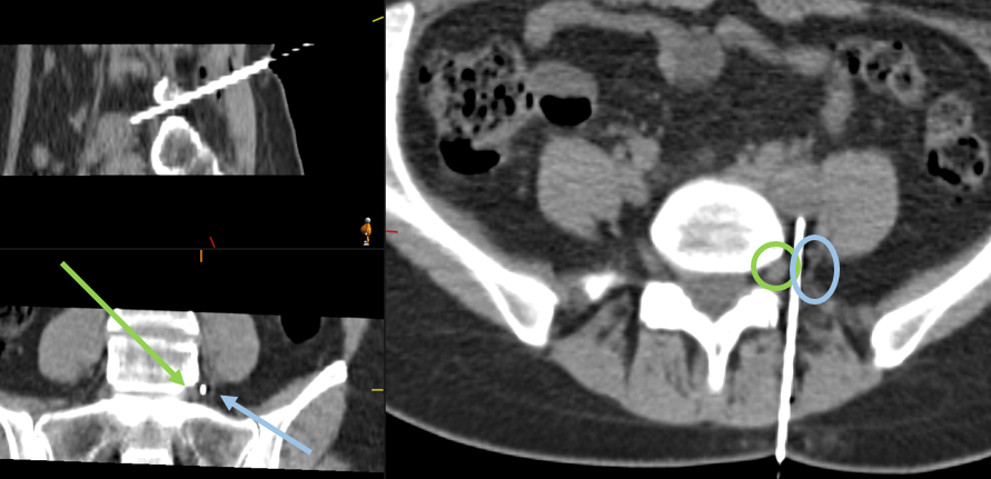
30F. What is your sound diagnosis?
Case Courtesy of Dr. Malak Itani, MD, Mallinckrodt
Trainees: Check the last image for details on how to send your answers.
#ultrasound #RadRes Malak Itani @MIRImaging


Don't miss this excellent case coauthored by colleagues from Mallinckrodt Institute of Radiology in the new issue of RadioGraphics


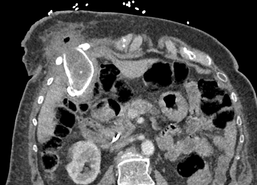
Ever seen a gallstone extending to an intercostal location?
Cholecystitis with body wall abscess!!!
Note: history is more complex with prior percutaneous cholecystostomy tube that was internalized..
#meded #radiology #abdrad #abdomen #imaging SAR DFP Benign Biliary Pathology


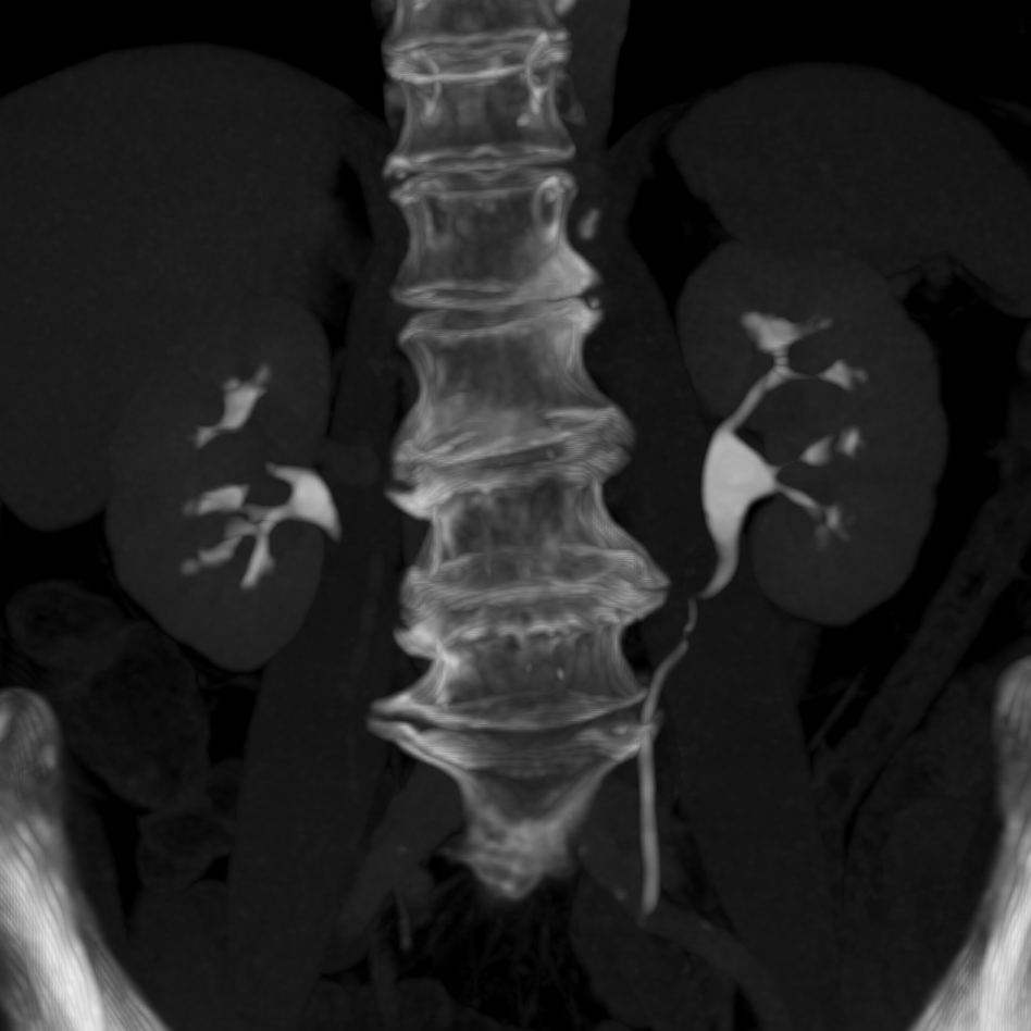
70 yo woman with hematuria, what's your diagnosis?
case from Mallinckrodt Institute of Radiology
#meded #radiology #abdrad #urology #nephrology

Multimodality Imaging in Metabolic Syndrome: State-of-the-Art Review
Kalisz K et al.
Multisystem Radiology
doi.org/10.1148/rg.230…
PR_CVRad
Sudhakar Venkatesh MD (ಸುಧಾಕರ್ ವೆಂಕಟೆೇಶ್)
Malak Itani
Amit Agarwal
#RGphx
7/12


Great representation from the Mallinckrodt Institute of Radiology abdomen section for this year's RadioGraphics Editor's Recognition Awards. Congrats David Ballard, Mark Hoegger, Dennis Balfe, Tyler Fraum, Hunter Lanier, Daniel Ludwig, and Jim Raptis.
pubs.rsna.org/doi/10.1148/rg…


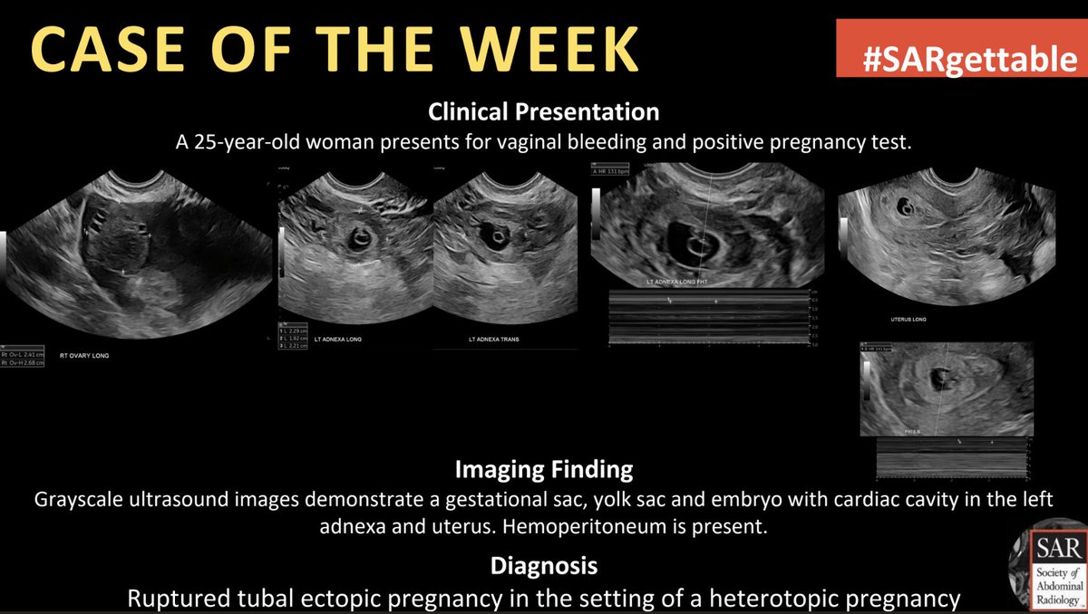
The answer to last week's #SARgettable Case of the Week is: Ruptured tubal ectopic pregnancy in the setting of a heterotopic pregnancy. Thanks for playing!


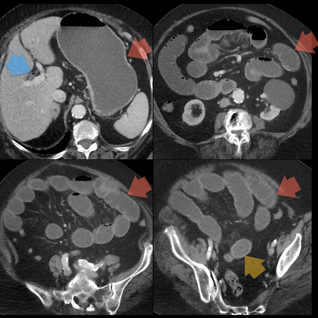
80-year-old female with pneumobilia (blue) + SBO (red) + large obstructing gallstone in the distal ileum (yellow) = Rigler's triad in gallstone ileus! Another Abdominal Radiology favorite! Brigham and Women's Radiology @AURtweet FOAMrad SAR Resident and Fellow Section Future Radiology Residents Harvard Macy CBR #MedEd
