Marcos Lepe, MD
@MarcosLepeMD
Pathologist at @harvardmed I @BIDMCpath | T/RT ≠ medical advice | Tweets = my opinion | #GUpath #Gynpath #Cytopath |
ID:45798931
https://www.marcoslepe.com 09-06-2009 09:00:30
7,5K Tweets
6,0K Followers
584 Following


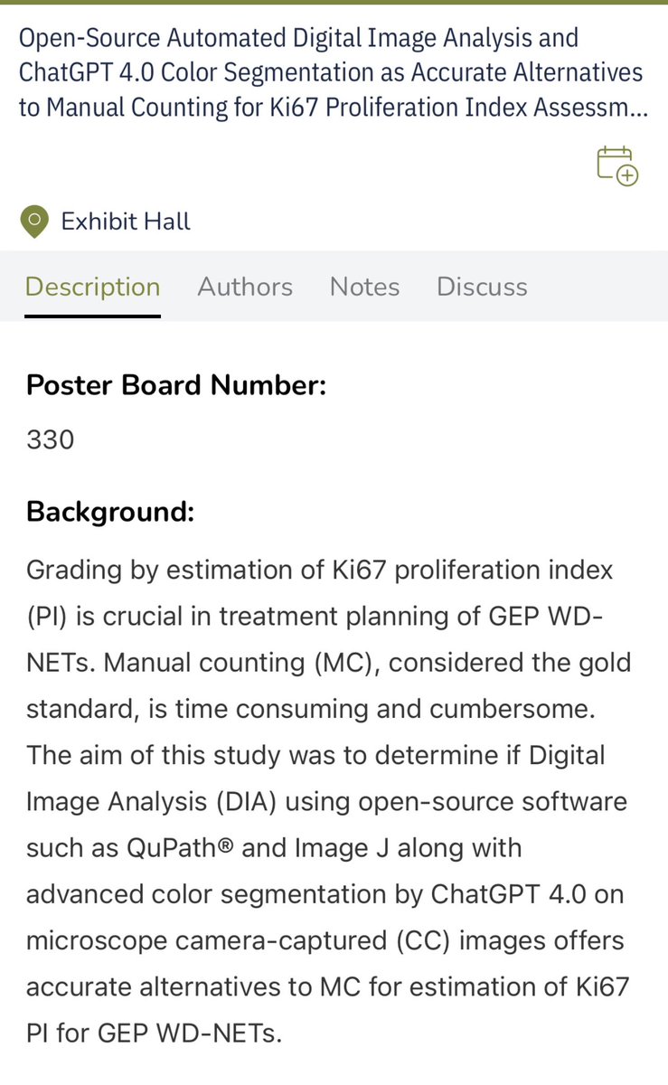
Congrats to BIDMC Pathology Nandan Padmanabha for winning the best abstract award from AIPNA!
You can check out his work today at the Stowell Orbison poster award session, poster #330
Laura Collins Vikram Deshpande Marcos Lepe, MD Liza Quintana, MD Michiya Nishino, MD, PhD

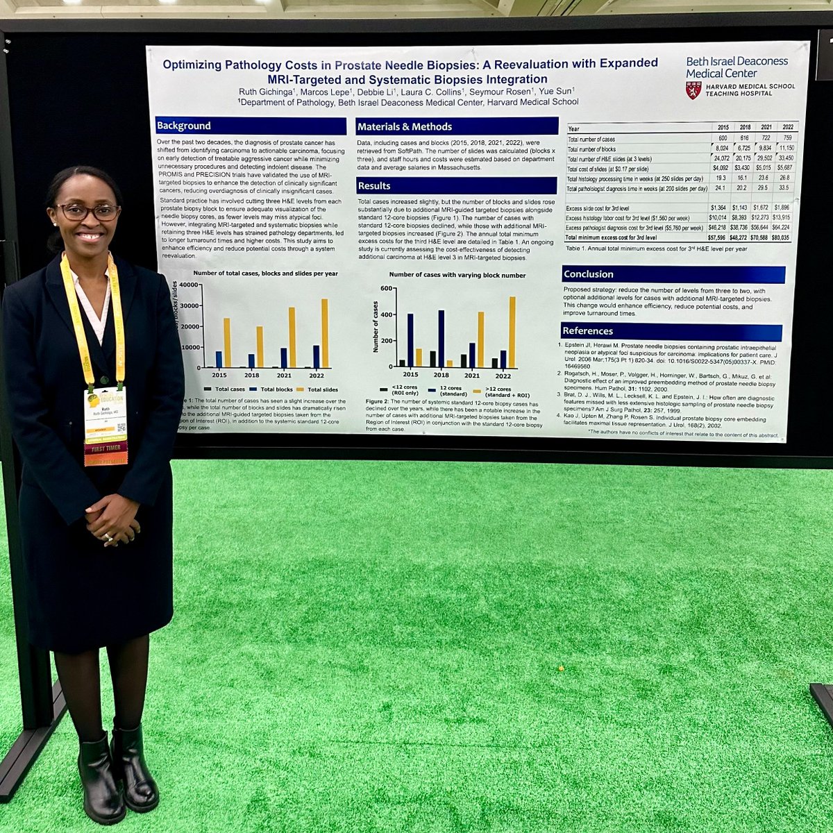
▶️Dr R. Gichinga's poster presentation at #USCAP2024
'Optimizing pathology costs in prostate needle biopsies: a reevaluation with expanded MRI-targeted and systematic biopsies integration'
Coauthors: Laura Collins, Marcos Lepe, MD, Dr S. Rosen, and Dr Y. Sun
So proud of our


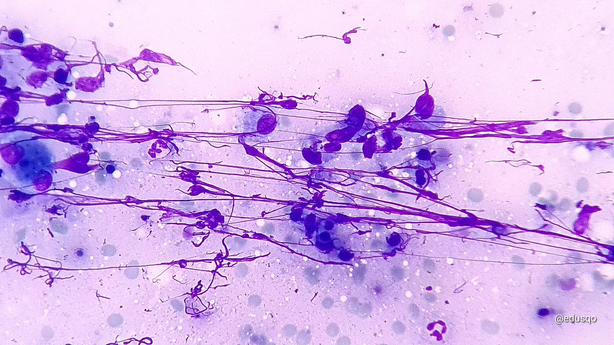
In a minority :( And in a bad company 🔬😧👻.
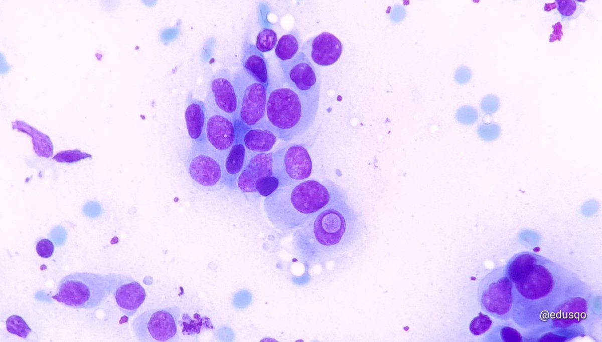
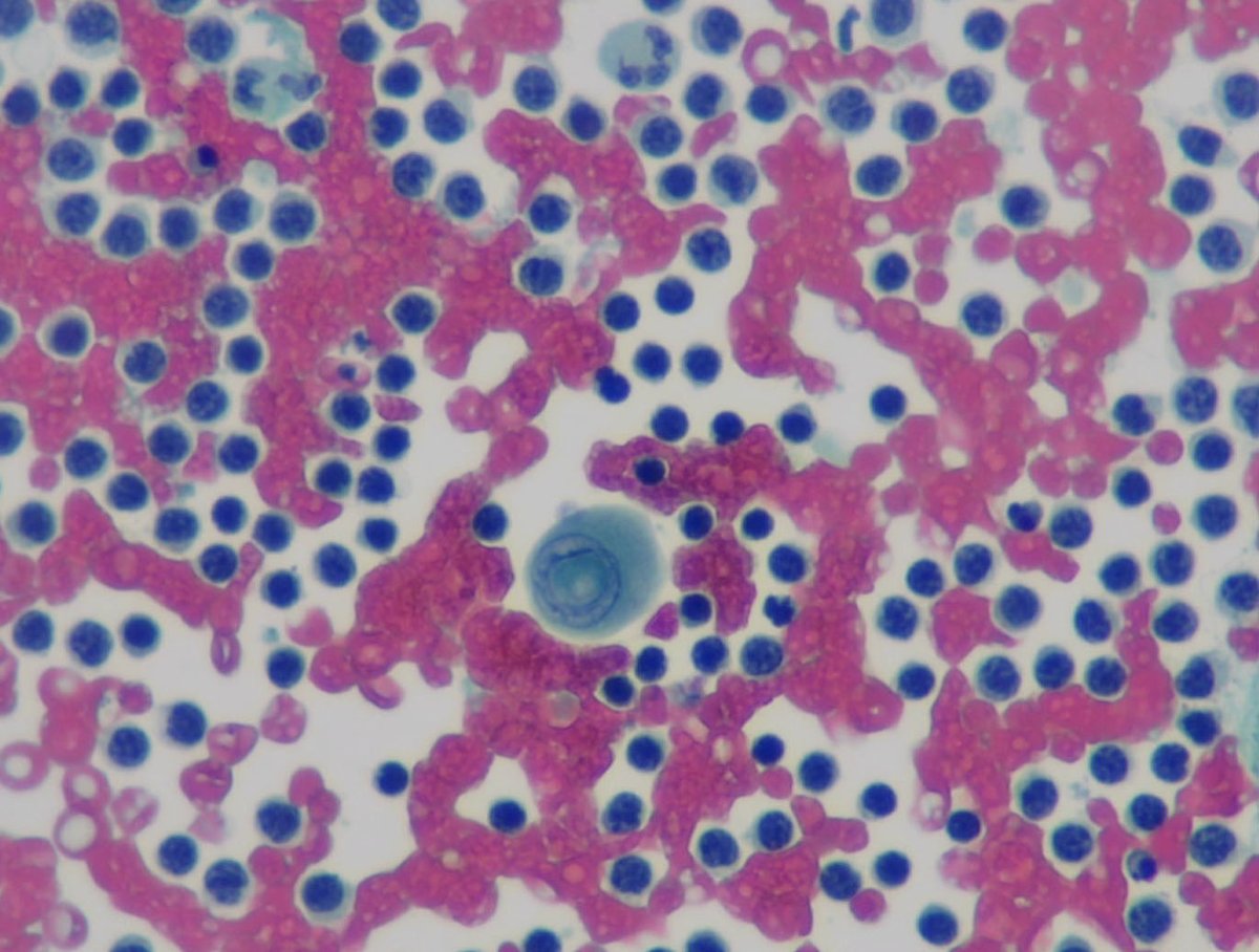
Liver mets of a PDAC Young EFCS Sociedad Española de Citología gulcin guler simsek, MD Quique Revilla
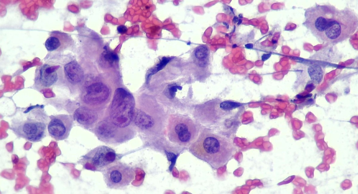


Small things matter. As Syed Z. Ali says, you can run but can't escape a cytopathologist's trained eyes. Papillary thyroid carcinoma in a lymph node #cytopath
Sociedad Española de Citología EFCS


Moving forward! Core Needle Biopsy on #FioNA ✅️.
Great performance of Elena Otón González, a fabulous SERVICIO DE RADIOLOGÍA HOSPITAL MORALES MESEGUER
Radiology resident.
#USFioNA #CNB #PatientSafety
Avanzando! Biopsia con Aguja Gruesa guiada ecográficamente de modo seguro. Entrenamiento con simulación.
#SegPac #BAG



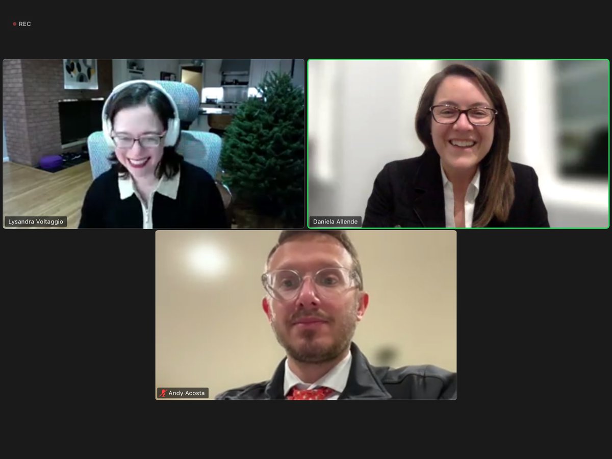
Ya estamos en vivo con la Dra. Lysandra Voltaggio presentado su clase sobre 'Patología Gastrointestinal'
#cursoLAPF #LAPF #Patologia
Daniela Allende, MD MBA Andres Martin Acosta (Andy) Lysandra Voltaggio, MD


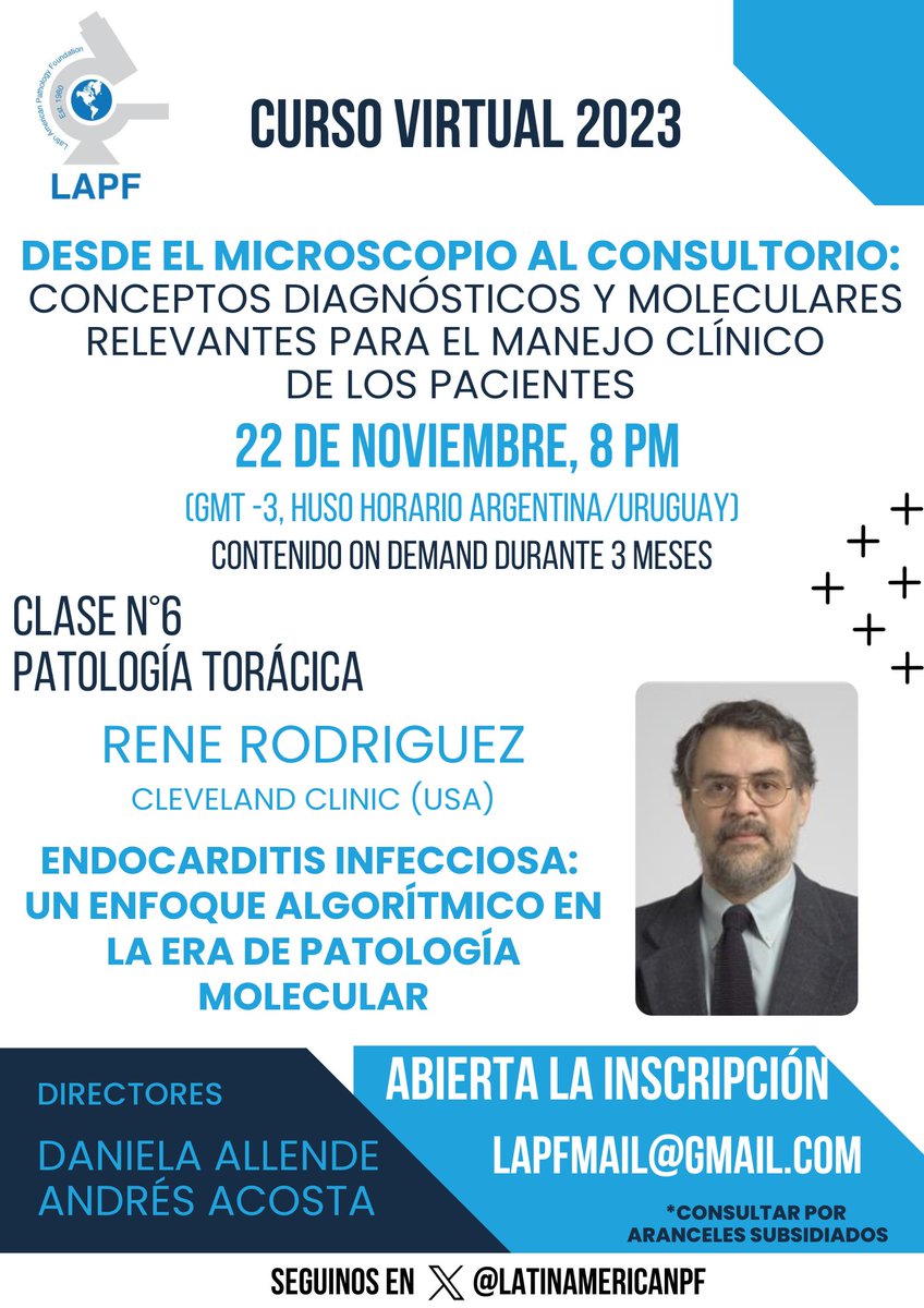
⚠️Recordatorio, hoy 8 PM (Argentina/Uruguay)
📍Clase N°6: Patología Torácica. Dr. René Rodríguez
Andres Martin Acosta (Andy) Daniela Allende, MD MBA Carlos Parra-Herran MD Andres Matoso Marcos Lepe, MD Fabiola Medeiros, MD
#cursovirtual #LAPF #patologíatorácica

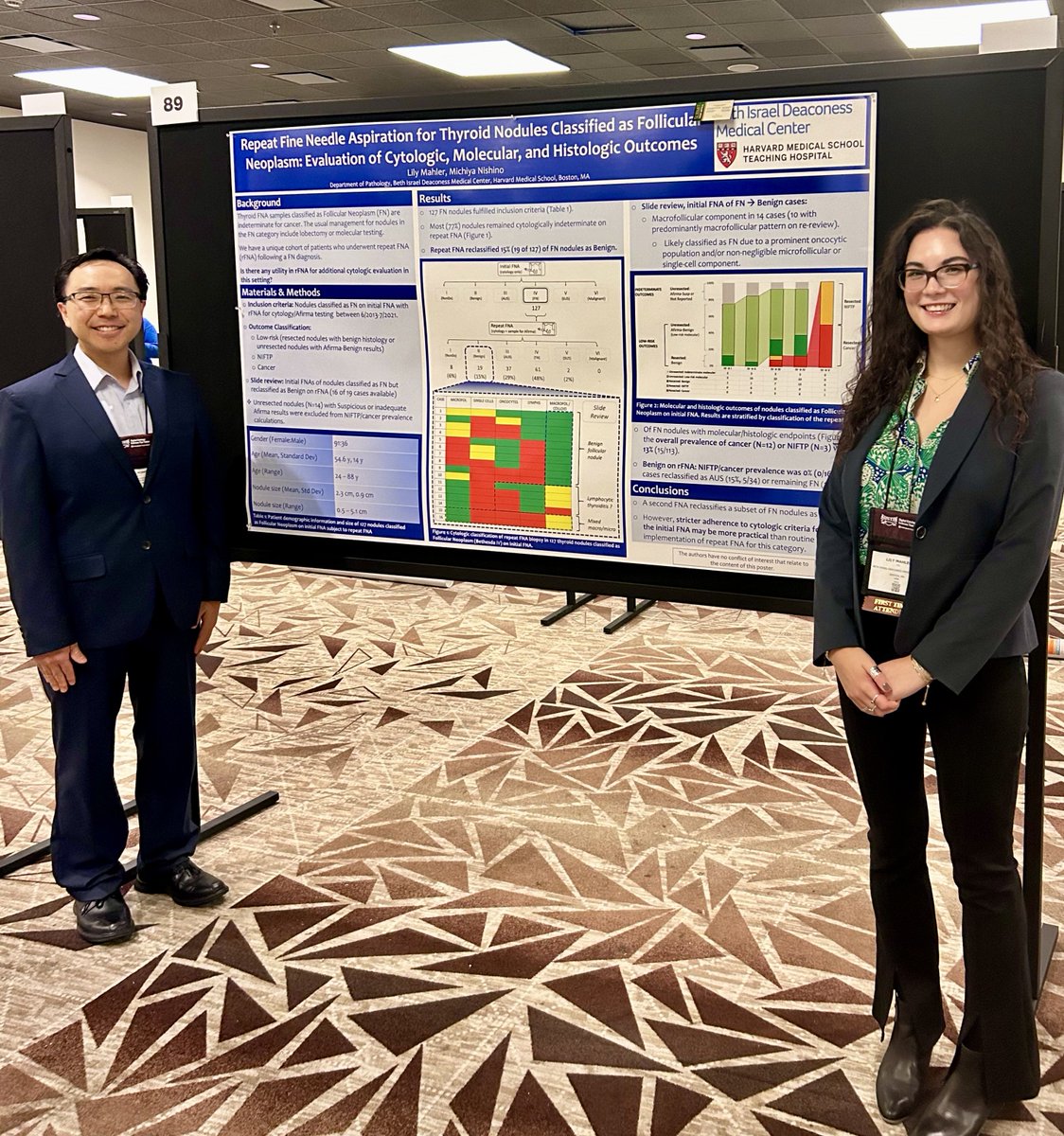
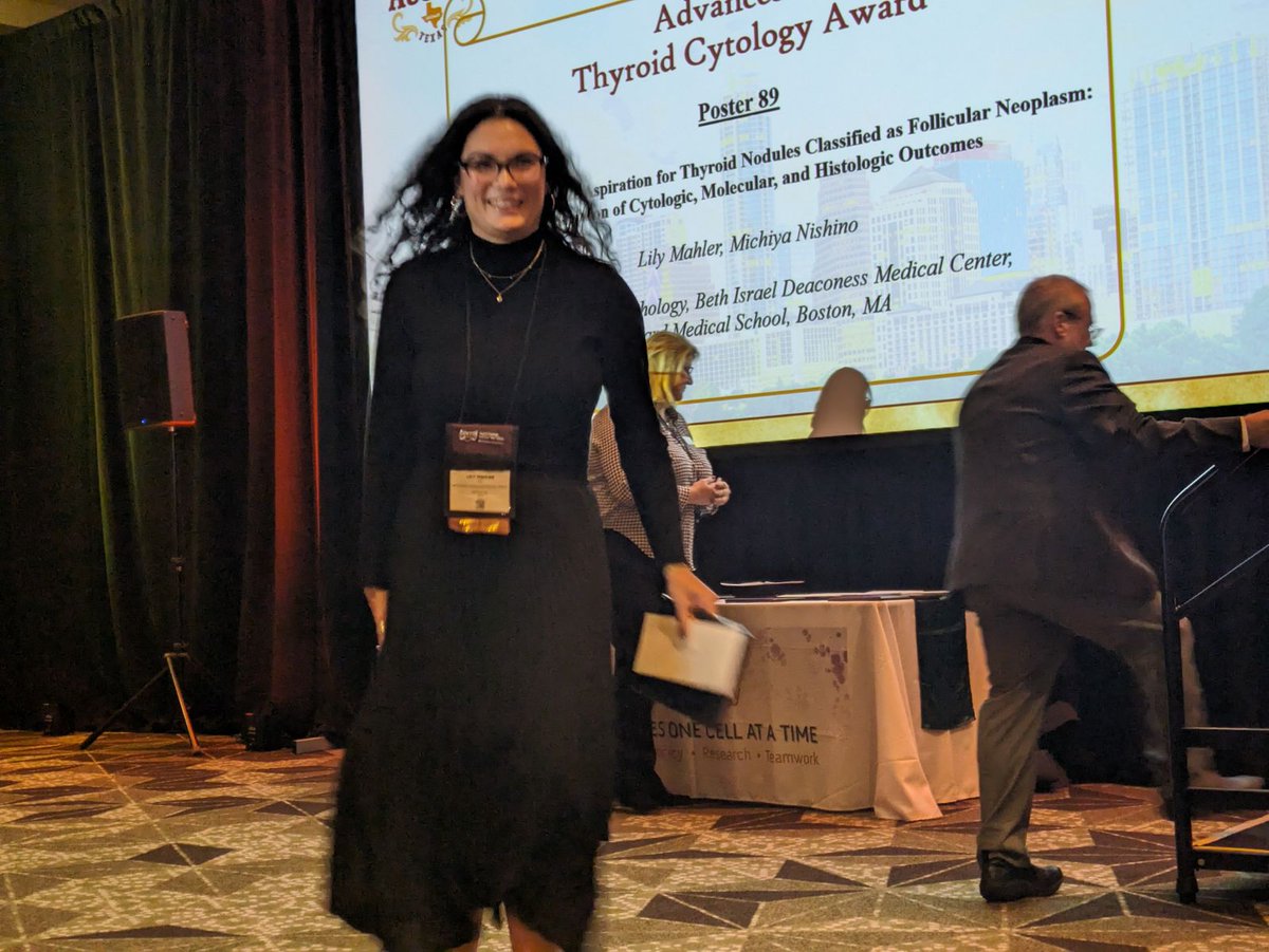
🔸🔶 Congratulations Lily Mahler, M.D. !!!🔶🔸
On receiving the Advances in Thyroid Cytology award at ASC's annual meeting!
Cytopathology.org #cytology #cytopathology #ASCyto23

⚜️BIDMC Pathology at #ASCyto23 ⚜️
Drs L. Mahler and M. Nishino next to their poster on 'Repeat FNA for Thyroid Nodules Classified as Follicular Neoplasm: Evaluation of Cytologic, Molecular and Histologic Outcomes'
At Cytopathology.org 's Annual Meeting!
BIDMC Pathology represent!!!
