Daniel Kowal, MD, RMSK | Radquarters(@Radquarters) 's Twitter Profileg
Daniel Kowal, MD, RMSK | Radquarters

@Radquarters

Radiologist & teacher: Body imaging, ultrasound, MSK radiology & more. Watch 📽️ Lectures: https://t.co/RrUYKt4dVP

ID:1094564487800197120

linkhttps://radquarters.com/ calendar_today10-02-2019 11:51:24

327 Tweets

5,8K Followers

372 Following

Daniel Kowal, MD, RMSK | Radquarters(@Radquarters) 's Twitter Profile Photo

Mercedes sign of gas-containing gallstones. Gallstone fissures may contain nitrogen gas yielding a triradiate lucent pattern. When present, aids in CT visualization of noncalcified gallstones. Not to be confused with emphysematous cholecystitis.

Mercedes sign of gas-containing gallstones. Gallstone fissures may contain nitrogen gas yielding a triradiate lucent pattern. When present, aids in CT visualization of noncalcified gallstones. Not to be confused with emphysematous cholecystitis. #FOAMrad #radres #medstudent
account_circle
Daniel Kowal, MD, RMSK | Radquarters(@Radquarters) 's Twitter Profile Photo

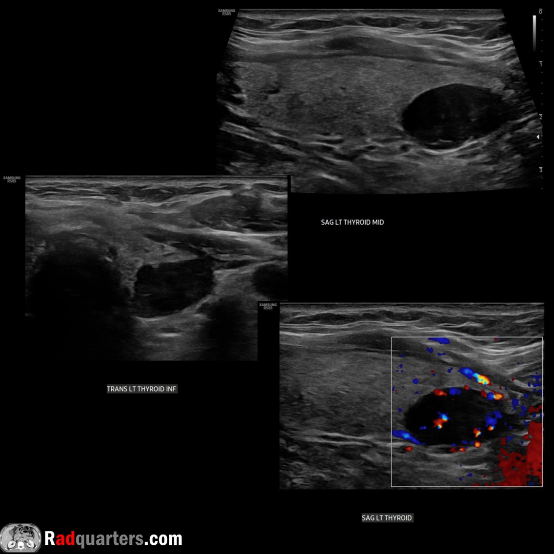
Large parathyroid adenoma. Usually oval/bean-shaped, but larger adenomas can be multilobulated. “Polar vessel” sign = Enlarged feeding artery or draining vein terminating @ parathyroid adenoma. Watch📽️ to learn more: bit.ly/pt-adenoma
Boston Imaging Samsung Healthcare

Large parathyroid adenoma. Usually oval/bean-shaped, but larger adenomas can be multilobulated. “Polar vessel” sign = Enlarged feeding artery or draining vein terminating @ parathyroid adenoma. Watch📽️ to learn more: bit.ly/pt-adenoma @BostonImaging @SamsungHealth #FOAMrad
account_circle
Daniel Kowal, MD, RMSK | Radquarters(@Radquarters) 's Twitter Profile Photo

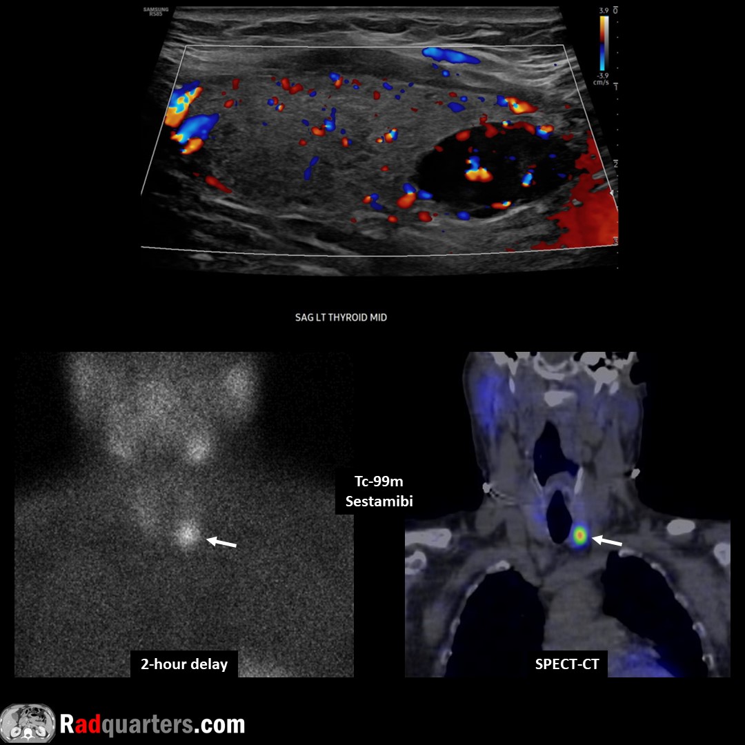
Parathyroid adenoma. Tc-99m sestamibi: Radiotracer initially taken up by both thyroid & parathyroid tissue but washes out more rapidly from thyroid & persists in adenoma on delayed images. Watch📽️ to learn more: bit.ly/pt-adenoma

Boston Imaging Samsung Healthcare

Parathyroid adenoma. Tc-99m sestamibi: Radiotracer initially taken up by both thyroid & parathyroid tissue but washes out more rapidly from thyroid & persists in adenoma on delayed images. Watch📽️ to learn more: bit.ly/pt-adenoma @BostonImaging @SamsungHealth #FOAMrad
account_circle
Daniel Kowal, MD, RMSK | Radquarters(@Radquarters) 's Twitter Profile Photo

Parathyroid adenoma. Most common cause of primary hyperPTH. Solid, homogeneous & very hypoechoic. Oval/bean-shaped, long axis craniocaudal. Hypervascular. Bright line separates from thyroid. Watch📽️ to learn more: bit.ly/pt-adenoma

Boston Imaging Samsung Healthcare

Parathyroid adenoma. Most common cause of primary hyperPTH. Solid, homogeneous & very hypoechoic. Oval/bean-shaped, long axis craniocaudal. Hypervascular. Bright line separates from thyroid. Watch📽️ to learn more: bit.ly/pt-adenoma @BostonImaging @SamsungHealth #FOAMrad
account_circle
Daniel Kowal, MD, RMSK | Radquarters(@Radquarters) 's Twitter Profile Photo

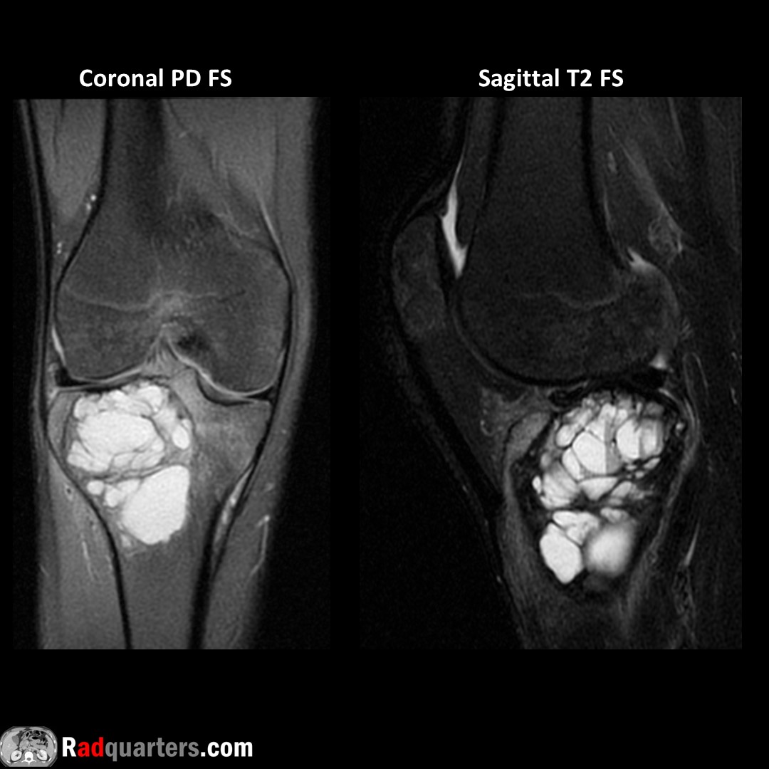
Giant cell tumor of proximal tibia. Eccentrically located at end of long bones (epiphysis + metaphyseal extension). Closed growth plates. Ages 20-40. +/- fluid-fluid levels, can co-exist with aneurysmal bone cyst. Rarely malignant but locally aggressive.

Giant cell tumor of proximal tibia. Eccentrically located at end of long bones (epiphysis + metaphyseal extension). Closed growth plates. Ages 20-40. +/- fluid-fluid levels, can co-exist with aneurysmal bone cyst. Rarely malignant but locally aggressive. #FOAMrad #radres #mskrad
account_circle
Daniel Kowal, MD, RMSK | Radquarters(@Radquarters) 's Twitter Profile Photo

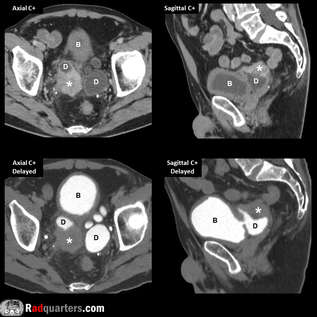
Urothelial carcinoma (*) arising in a bladder (B) diverticulum (D). Bladder most common site for urothelial carcinoma (AKA transitional cell carcinoma), but only rarely seen in diverticula. Bladder diverticula can also be complicated with infection or calculi.

Urothelial carcinoma (*) arising in a bladder (B) diverticulum (D). Bladder most common site for urothelial carcinoma (AKA transitional cell carcinoma), but only rarely seen in diverticula. Bladder diverticula can also be complicated with infection or calculi. #FOAMrad #radres
account_circle
Daniel Kowal, MD, RMSK | Radquarters(@Radquarters) 's Twitter Profile Photo

Juvenile recurrent parotitis. Multiple nonuniform hypoechoic foci scattered throughout gland with central calcifications (arrows). Rare but 2nd most common cause of parotitis in childhood after mumps. Watch📽️ to learn more: bit.ly/rq-parotitis

Boston Imaging Samsung Healthcare
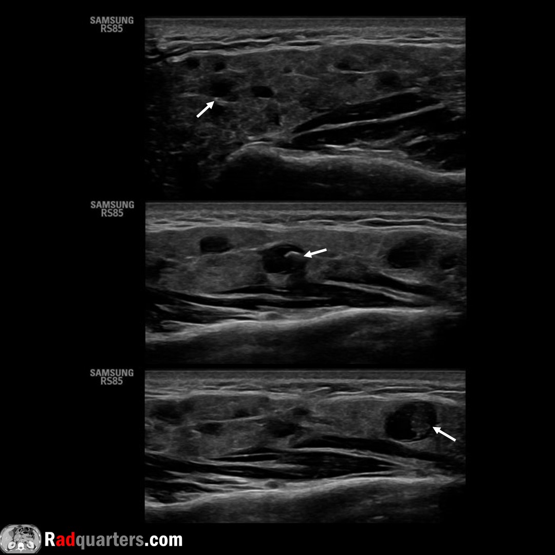
Juvenile recurrent parotitis. Multiple nonuniform hypoechoic foci scattered throughout gland with central calcifications (arrows). Rare but 2nd most common cause of parotitis in childhood after mumps. Watch📽️ to learn more: bit.ly/rq-parotitis @BostonImaging @SamsungHealth
account_circle
Daniel Kowal, MD, RMSK | Radquarters(@Radquarters) 's Twitter Profile Photo

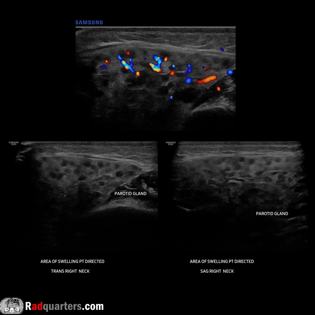
Pomegranate sign of acute parotitis. Uniform anechoic foci scattered throughout the gland. Similar to pseudonodular “giraffe” ultrasound pattern that can be seen in Hashimoto’s thyroiditis. Watch📽️ to learn more: bit.ly/rq-parotitis
Boston Imaging Samsung Healthcare

Pomegranate sign of acute parotitis. Uniform anechoic foci scattered throughout the gland. Similar to pseudonodular “giraffe” ultrasound pattern that can be seen in Hashimoto’s thyroiditis. Watch📽️ to learn more: bit.ly/rq-parotitis @BostonImaging @SamsungHealth #FOAMrad
account_circle
Daniel Kowal, MD, RMSK | Radquarters(@Radquarters) 's Twitter Profile Photo

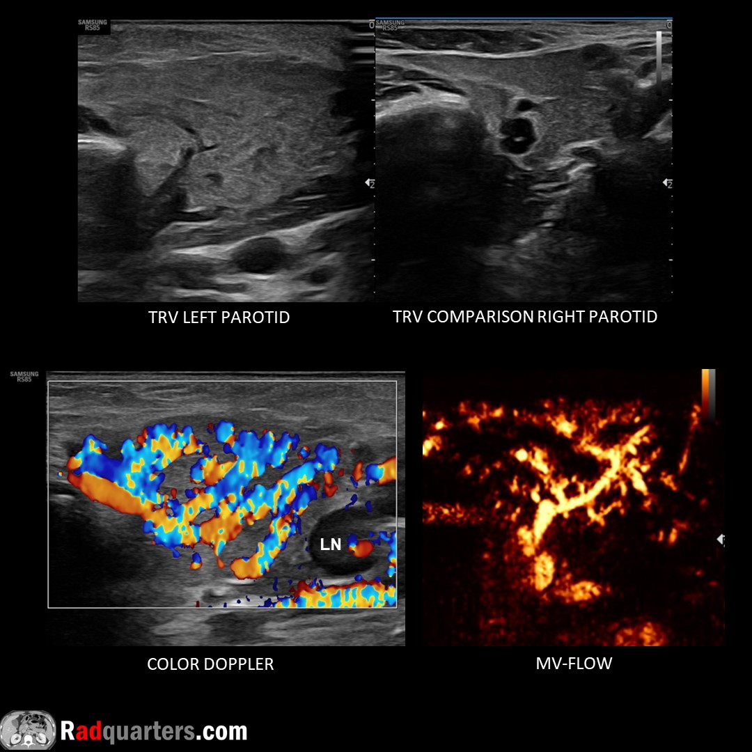
Acute left parotitis. 2 y.o. with enlarged, heterogeneous, hyperemic left parotid gland with adjacent lymphadenopathy (LN). Normal right gland. In children, most common cause viral (mumps). Watch📽️ to learn more: bit.ly/rq-parotitis

Boston Imaging Samsung Healthcare

Acute left parotitis. 2 y.o. with enlarged, heterogeneous, hyperemic left parotid gland with adjacent lymphadenopathy (LN). Normal right gland. In children, most common cause viral (mumps). Watch📽️ to learn more: bit.ly/rq-parotitis @BostonImaging @SamsungHealth #FOAMrad
account_circle
Daniel Kowal, MD, RMSK | Radquarters(@Radquarters) 's Twitter Profile Photo

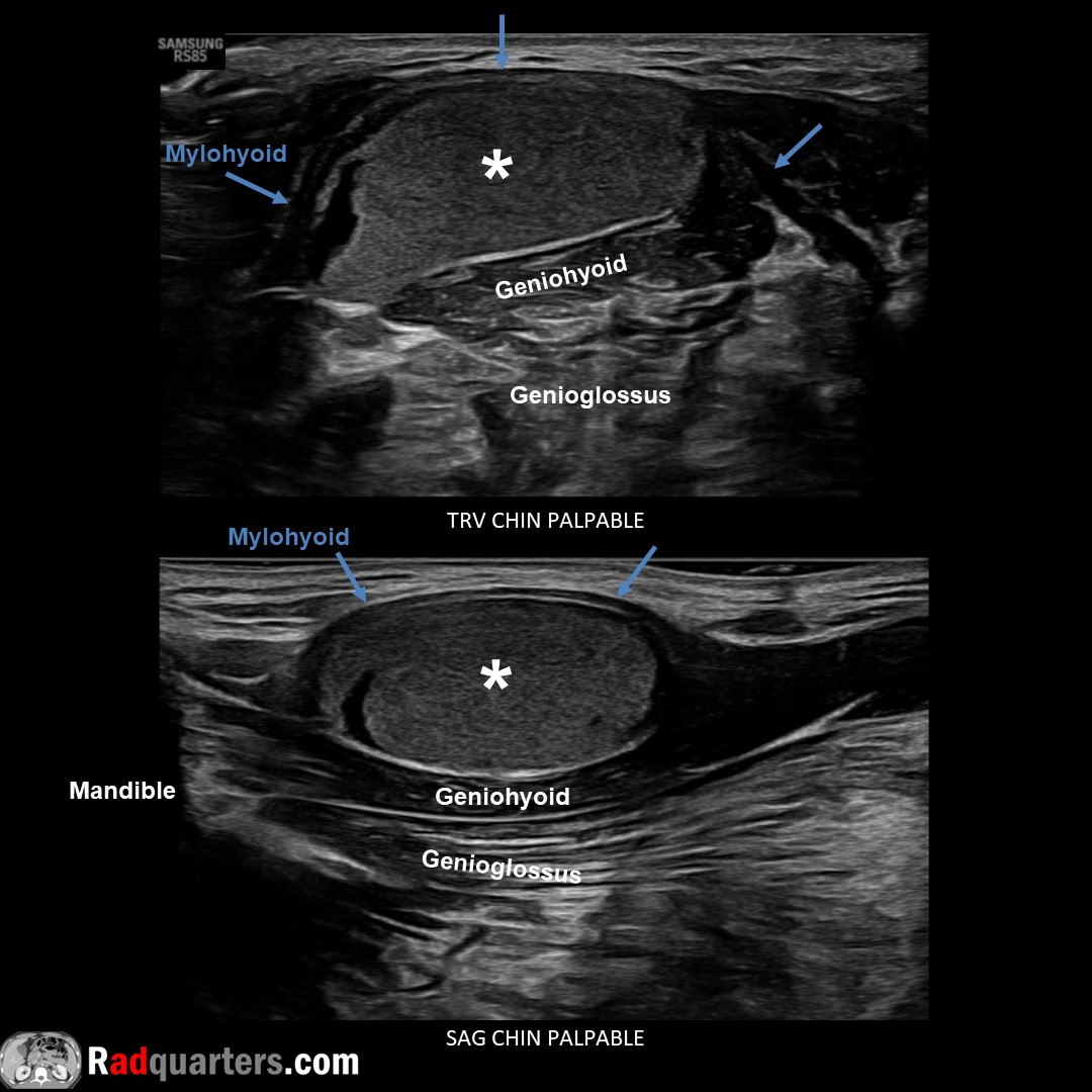
Sublingual dermoid cyst. Rare, benign, squamous epithelial lining & contains skin appendages. Sublingual space bounded medially by genioglossus/geniohyoid muscles, inferolaterally by mylohyoid. Watch📽️ to learn more: bit.ly/subderm
Boston Imaging Samsung Healthcare

Sublingual dermoid cyst. Rare, benign, squamous epithelial lining & contains skin appendages. Sublingual space bounded medially by genioglossus/geniohyoid muscles, inferolaterally by mylohyoid. Watch📽️ to learn more: bit.ly/subderm @BostonImaging @SamsungHealth #FOAMrad
account_circle
Daniel Kowal, MD, RMSK | Radquarters(@Radquarters) 's Twitter Profile Photo

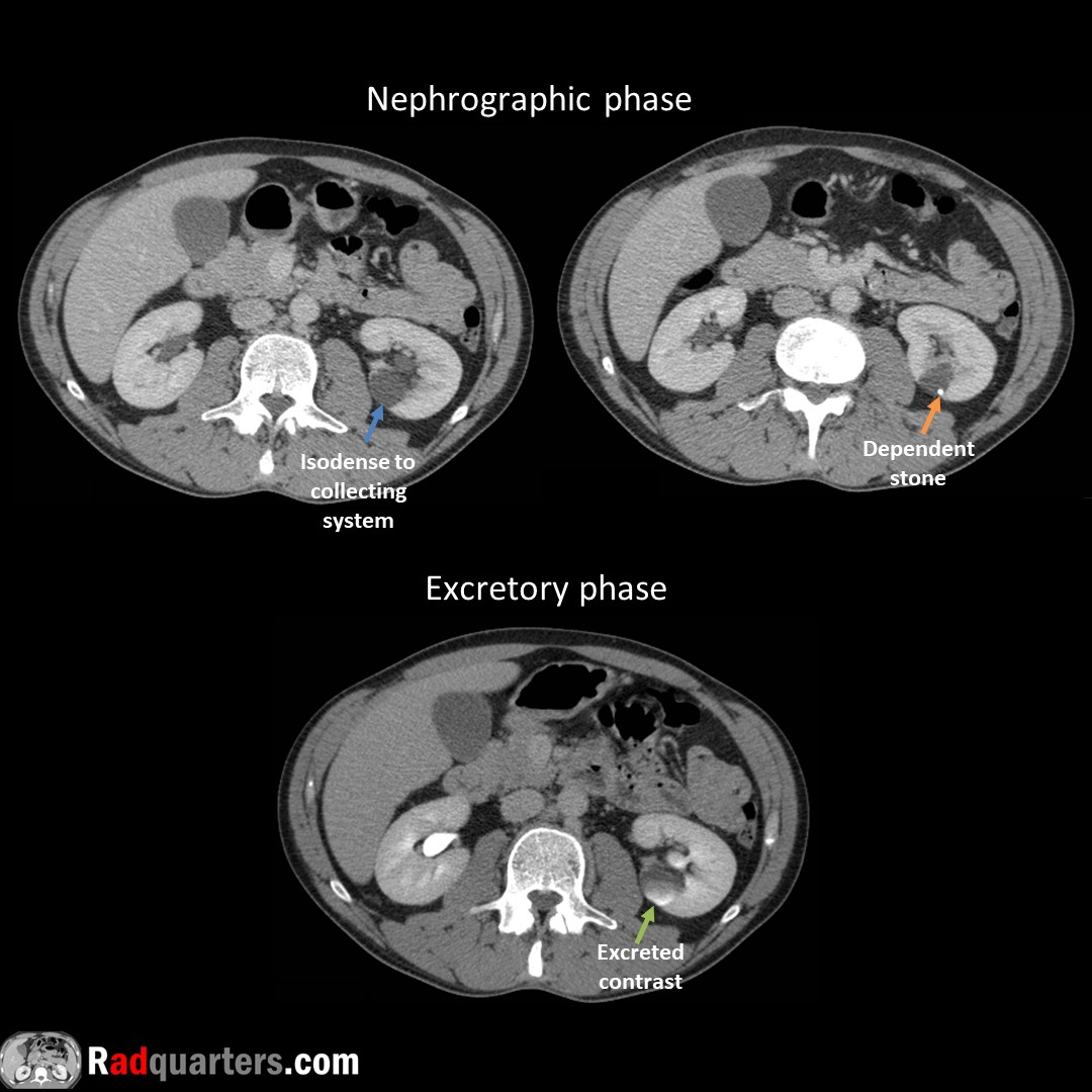
Calyceal diverticulum. Often asymptomatic. Can present with hematuria, stone formation or infection. Mimics cyst on conventional imaging, contains excreted contrast on excretory phase due to communication with collecting system.

Calyceal diverticulum. Often asymptomatic. Can present with hematuria, stone formation or infection. Mimics cyst on conventional imaging, contains excreted contrast on excretory phase due to communication with collecting system. #FOAMrad #FOAMed #radres #medstudent #radquarters
account_circle
Daniel Kowal, MD, RMSK | Radquarters(@Radquarters) 's Twitter Profile Photo

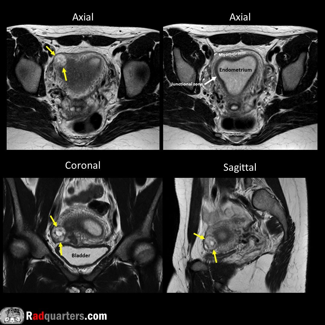
Interstitial ectopic pregnancy on T2W MRI. Pregnancy within proximal fallopian tube surrounded by uterine wall. Higher risk of rupture compared to the more common tubal ectopic pregnancy. Sac located eccentric to junctional zone with thin (<5 mm) overlying myometrium.

Interstitial ectopic pregnancy on T2W MRI. Pregnancy within proximal fallopian tube surrounded by uterine wall. Higher risk of rupture compared to the more common tubal ectopic pregnancy. Sac located eccentric to junctional zone with thin (<5 mm) overlying myometrium. #FOAMrad
account_circle
Daniel Kowal, MD, RMSK | Radquarters(@Radquarters) 's Twitter Profile Photo

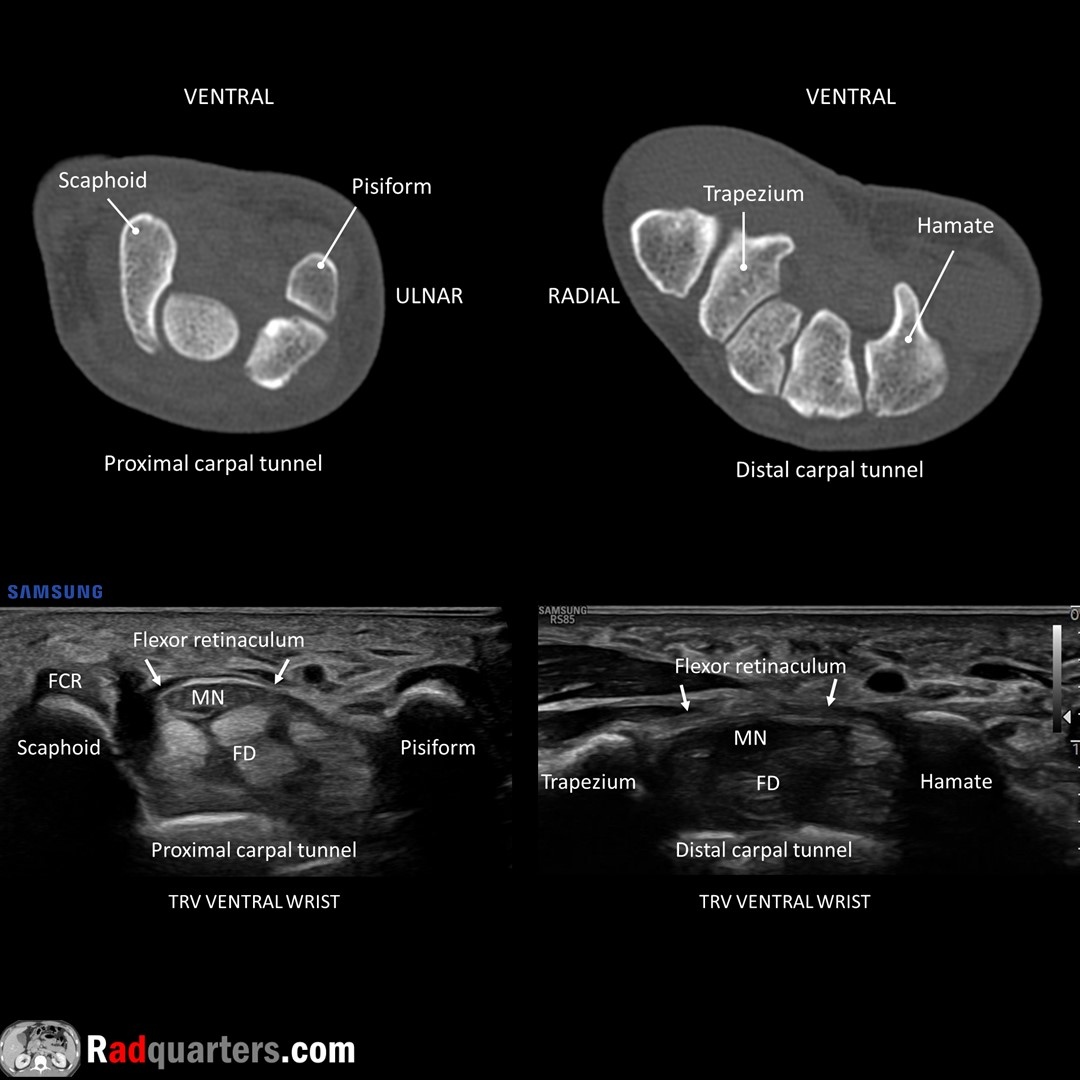
Carpal tunnel anatomy. Median nerve (MN) deep to flexor retinaculum, anterior to flexor digitorum superficialis & profundus tendons (FD). Flexor carpi radialis tendon (FCR) anterior to scaphoid. Watch📽️ to learn more: bit.ly/rq-cts
Boston Imaging Samsung Healthcare

Carpal tunnel anatomy. Median nerve (MN) deep to flexor retinaculum, anterior to flexor digitorum superficialis & profundus tendons (FD). Flexor carpi radialis tendon (FCR) anterior to scaphoid. Watch📽️ to learn more: bit.ly/rq-cts @BostonImaging @SamsungHealth #FOAMrad
account_circle
Daniel Kowal, MD, RMSK | Radquarters(@Radquarters) 's Twitter Profile Photo

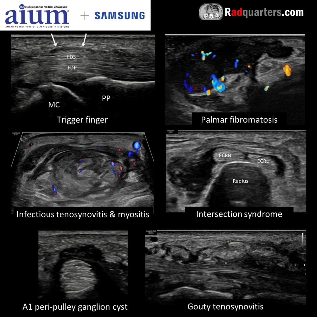
Join me for “Ultrasound of the Hand & Wrist: What the Surgeon Wants to Know,” a FREE webinar Wed,11/8/2023 at 1 PM EST hosted by the AIUM in conjunction w/ Samsung. Register here: bit.ly/wristwebinar, or go to aium.org. Boston Imaging AIUM Ultrasound

Join me for “Ultrasound of the Hand & Wrist: What the Surgeon Wants to Know,” a FREE webinar Wed,11/8/2023 at 1 PM EST hosted by the AIUM in conjunction w/ Samsung. Register here: bit.ly/wristwebinar, or go to aium.org. @BostonImaging @AIUMultrasound #mskrad
account_circle
Daniel Kowal, MD, RMSK | Radquarters(@Radquarters) 's Twitter Profile Photo

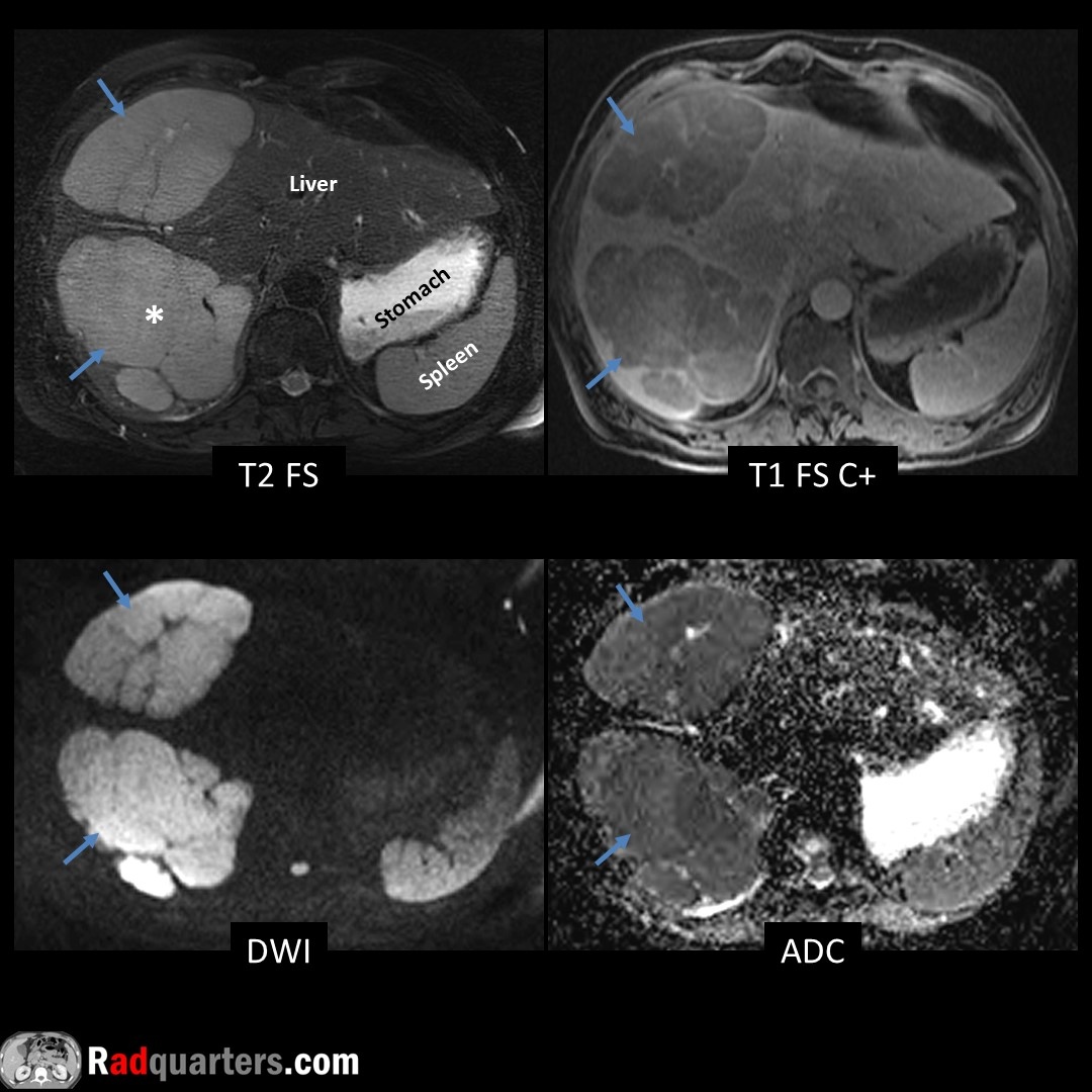
Multifocal primary hepatic lymphoma. Rare. Often solitary unlike diffusely infiltrating secondary lymphoma. T1 dark, T2 bright (relative to liver), hypoenhancing w/ faint rim enhancement. Marked restricted diffusion due to hypercellularity. Path = High-grade B-cell.

Multifocal primary hepatic lymphoma. Rare. Often solitary unlike diffusely infiltrating secondary lymphoma. T1 dark, T2 bright (relative to liver), hypoenhancing w/ faint rim enhancement. Marked restricted diffusion due to hypercellularity. Path = High-grade B-cell. #FOAMrad
account_circle
Daniel Kowal, MD, RMSK | Radquarters(@Radquarters) 's Twitter Profile Photo

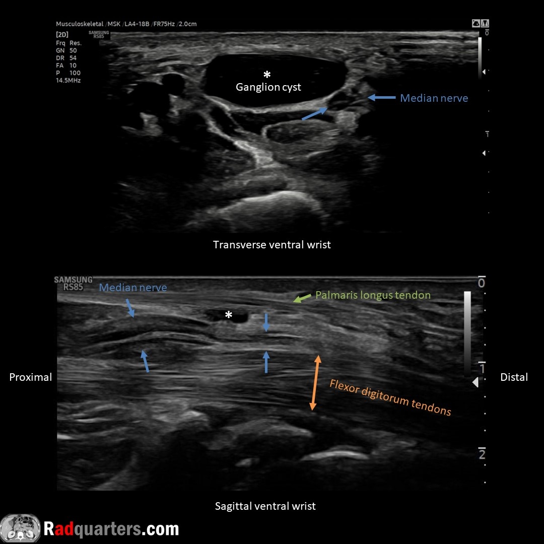
Ventral wrist ganglion cyst causing median nerve compression. MN identified by its fascicular, honeycomb configuration. Thickened nerve proximal to cyst, normal diameter distally = Notch sign. Watch📽️ to learn more: bit.ly/rq-ganglion
Boston Imaging Samsung Healthcare

Ventral wrist ganglion cyst causing median nerve compression. MN identified by its fascicular, honeycomb configuration. Thickened nerve proximal to cyst, normal diameter distally = Notch sign. Watch📽️ to learn more: bit.ly/rq-ganglion @BostonImaging @SamsungHealth #FOAMrad
account_circle
Daniel Kowal, MD, RMSK | Radquarters(@Radquarters) 's Twitter Profile Photo

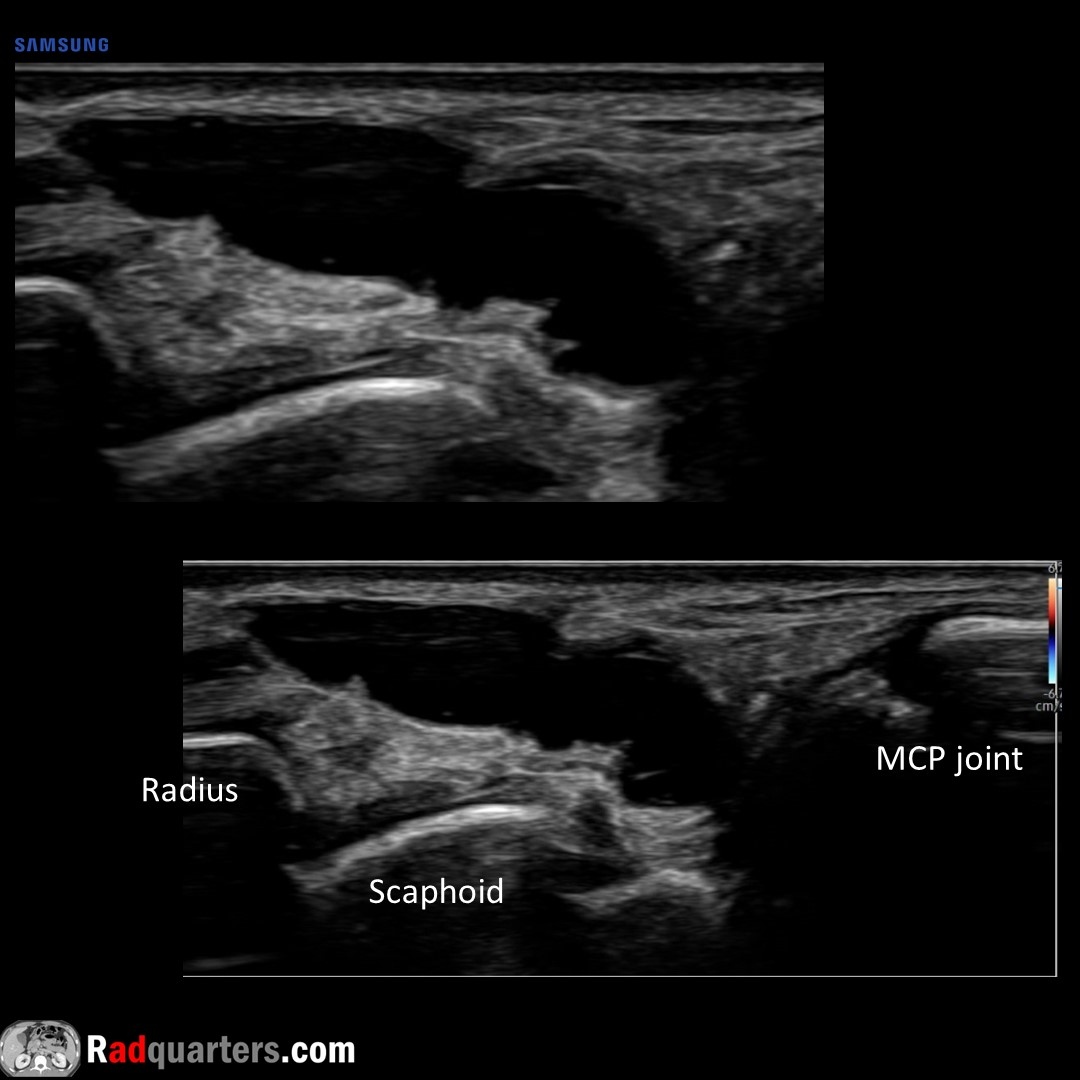
Dorsal wrist ganglion cyst. May see pedicle connecting to joint. Dorsal wrist most common location. Unlike joint recess fluid & bursal collections, will not collapse with compression.
Watch📽️ to learn more: bit.ly/rq-ganglion
Boston Imaging Samsung Healthcare

Dorsal wrist ganglion cyst. May see pedicle connecting to joint. Dorsal wrist most common location. Unlike joint recess fluid & bursal collections, will not collapse with compression. Watch📽️ to learn more: bit.ly/rq-ganglion @BostonImaging @SamsungHealth #FOAMrad
account_circle
Daniel Kowal, MD, RMSK | Radquarters(@Radquarters) 's Twitter Profile Photo

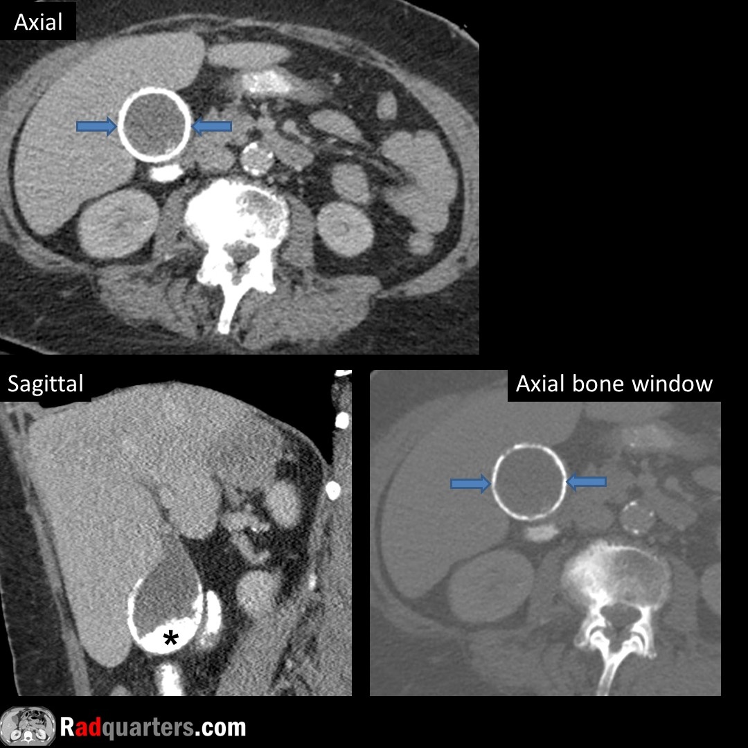
Porcelain gallbladder. Dense calcification outlining gallbladder wall (arrows). Most (90-95%) have associated gallstones (*). Originally thought to have a high association with GB adenocarcinoma, but more recent studies have shown a much weaker risk (<1% per year).

Porcelain gallbladder. Dense calcification outlining gallbladder wall (arrows). Most (90-95%) have associated gallstones (*). Originally thought to have a high association with GB adenocarcinoma, but more recent studies have shown a much weaker risk (<1% per year). #FOAMrad
account_circle
Daniel Kowal, MD, RMSK | Radquarters(@Radquarters) 's Twitter Profile Photo

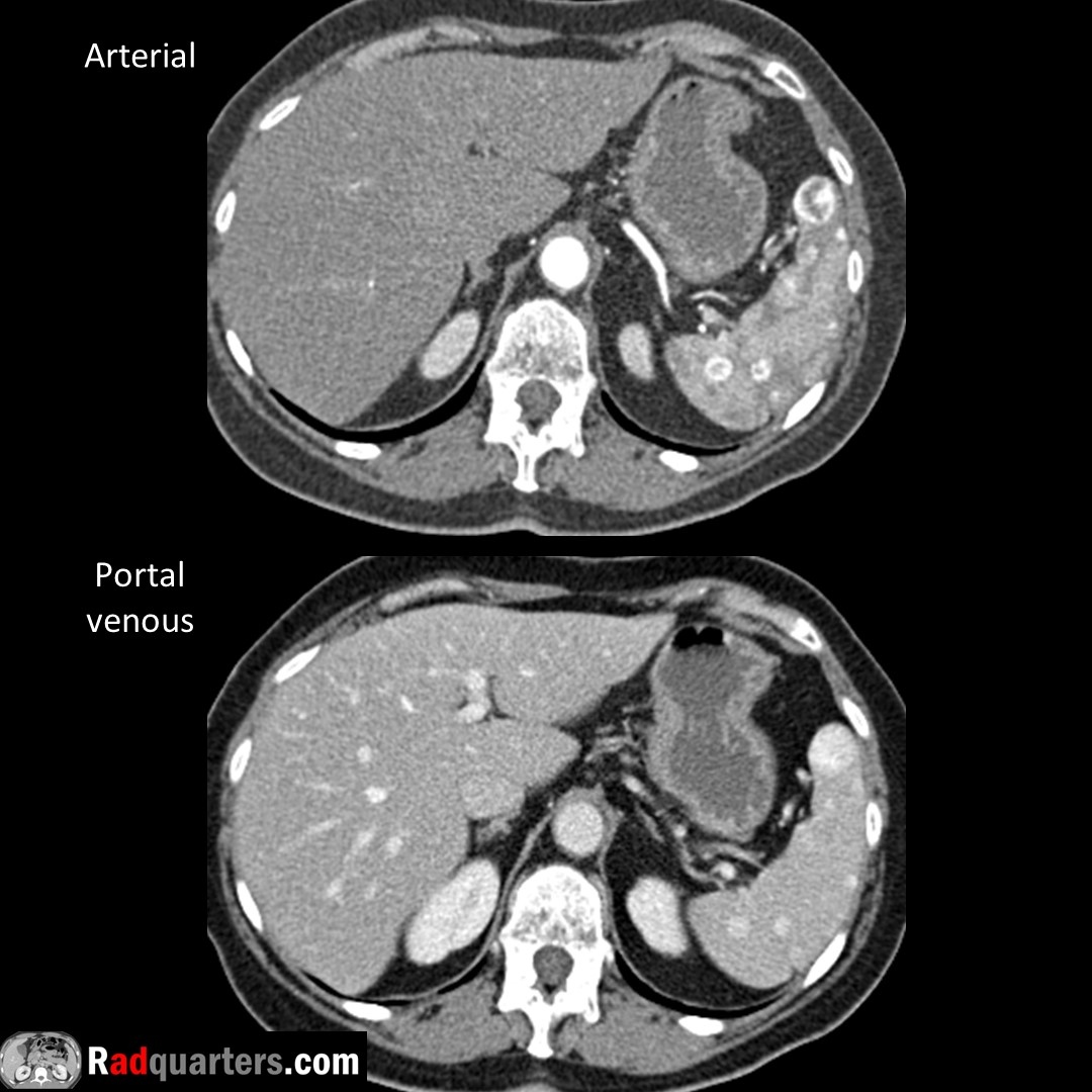
Multiple splenic hemangiomas. Most common benign neoplasm of spleen. Centripetal enhancement from periphery inward, uniform fill-in on delayed images = Similar enhancement to hepatic hemangiomas, except characteristic peripheral nodular pattern often absent.

Multiple splenic hemangiomas. Most common benign neoplasm of spleen. Centripetal enhancement from periphery inward, uniform fill-in on delayed images = Similar enhancement to hepatic hemangiomas, except characteristic peripheral nodular pattern often absent. #FOAMrad #radres
account_circle
Daniel Kowal, MD, RMSK | Radquarters(@Radquarters) 's Twitter Profile Photo

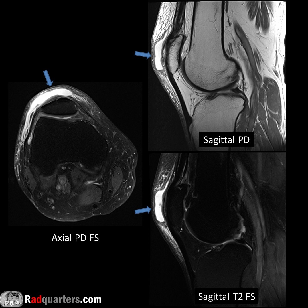
Prepatellar bursitis. Fluid accumulation anterior to patella in prepatellar bursa (arrows). Typically caused by direct trauma or repetitive kneeling. Less commonly caused by gout, RA, or infection. May see calcifications if chronic.

Prepatellar bursitis. Fluid accumulation anterior to patella in prepatellar bursa (arrows). Typically caused by direct trauma or repetitive kneeling. Less commonly caused by gout, RA, or infection. May see calcifications if chronic. #FOAMrad #mskrad #radres #medstudent
account_circle