Andy Moore(@aaandmoore) 's Twitter Profileg
Andy Moore

@aaandmoore

Cell biologist interested in organelles and how they move. Husband, dad, intermediate filament apologist, and postdoc in the @JLS_Lab at @HHMIJanelia.

ID:782222741651030020

linkhttps://scholar.google.com/citations?user=pSN-aH4AAAAJ&hl=en calendar_today01-10-2016 14:16:49

2,6K Tweets

7,5K Followers

3,2K Following

Andy Moore(@aaandmoore) 's Twitter Profile Photo

This is a movie of ER fission in which fragmented tubules seem to fully disconnect from the reticulum before re-fusing soon after. Red box=fission site, yellow arrow = free tubule, cyan circle = fusion site.

account_circle
Erika Holzbaur-Howland(@ErikaHolzbaur) 's Twitter Profile Photo

So proud of the new work out today from Liv Harding, Julia Riley, Elisabeth Holzer and Sascha Martens: Damaged mitochondria recruit the effector NEMO to activate NF-κB signaling: Molecular Cell cell.com/molecular-cell…

account_circle
Andy Moore(@aaandmoore) 's Twitter Profile Photo

This is a ~6h movie of U2OS cells migrating into a scratch. Magenta = labeled vimentin at time 0, green = newly translated vimentin.

account_circle
Eric Peterman(@errricpeterman) 's Twitter Profile Photo

not to be outdone by myself, i explanted and imaged zebrafish skin over 8 hours to watch cell motility on a tissue-wide scale.

we're watching mpeg1+ immune cells surveil the skin, while some react to a skin injury i inadvertently caused in the skin explant process (top middle)

account_circle
Eric Peterman(@errricpeterman) 's Twitter Profile Photo

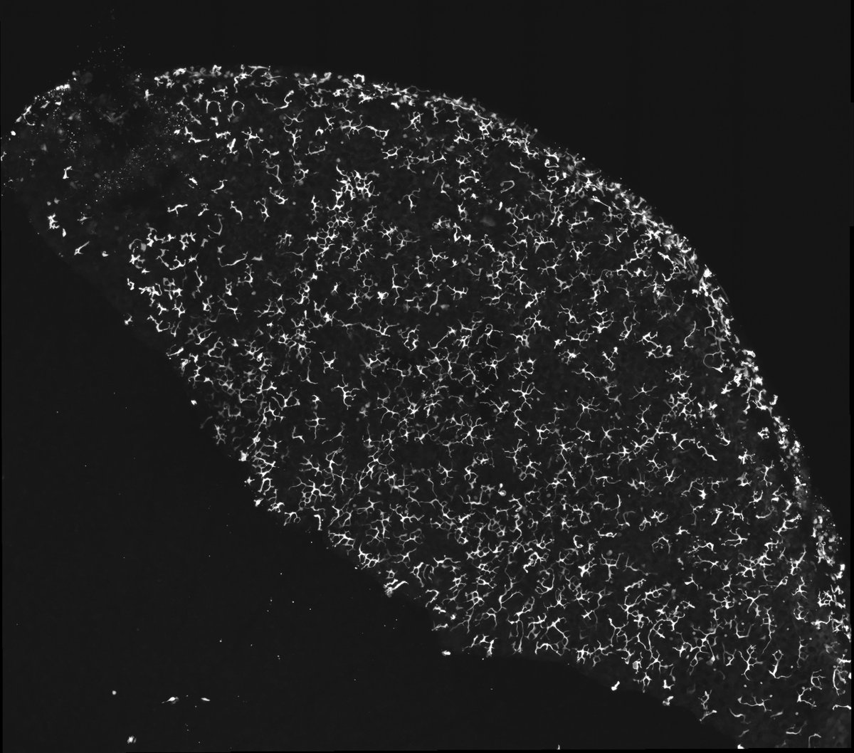
i should do stitched imaging more often, these are lovely (in my biased opinion)
mpeg1+ cells in zebrafish skin explant

i should do stitched imaging more often, these are lovely (in my biased opinion) mpeg1+ cells in zebrafish skin explant
account_circle
Dr. Adelita Mendoza(@AdelitaMendoza7) 's Twitter Profile Photo

Dr. Arnaldo Díaz Vázquez Me!! I’m Adelita and I study the role of lysosomes in zinc homeostasis using C.elegans and human models systems. I use advanced imaging, genetics, and molecular biology, and x-ray physics techniques in my work.

account_circle
Shekhar lab(@sshekhr) 's Twitter Profile Photo

📢(1/n) Please RT!

I am hiring a postdoc to work on exciting biophysics and biochemical projects in actin mechanobiology in my lab at Emory University, Atlanta, USA!

See lab website: shekharlab.org

account_circle
Simpson Lab @UWMedicine(@SimpsonLabUW) 's Twitter Profile Photo

Excited 🥳 to get official notice for 1st (non-transferred) NIAMS grant to UW Medicine (@SimpsonLabUW) 's Twitter Profile">Simpson Lab UW Medicine UW Medicine. Via this R03, we’ll use live microscopy of organoid human skin to learn how keratinocytes use to remodel/degrade organelles like ER to form the epidermal barrier.

Excited 🥳 to get official notice for 1st (non-transferred) @NIH_NIAMS grant to @SimpsonLabUW @UWMedicine. Via this R03, we’ll use live microscopy of organoid human skin to learn how keratinocytes use #autophagy to remodel/degrade organelles like ER to form the epidermal barrier.
account_circle
Andy Moore(@aaandmoore) 's Twitter Profile Photo

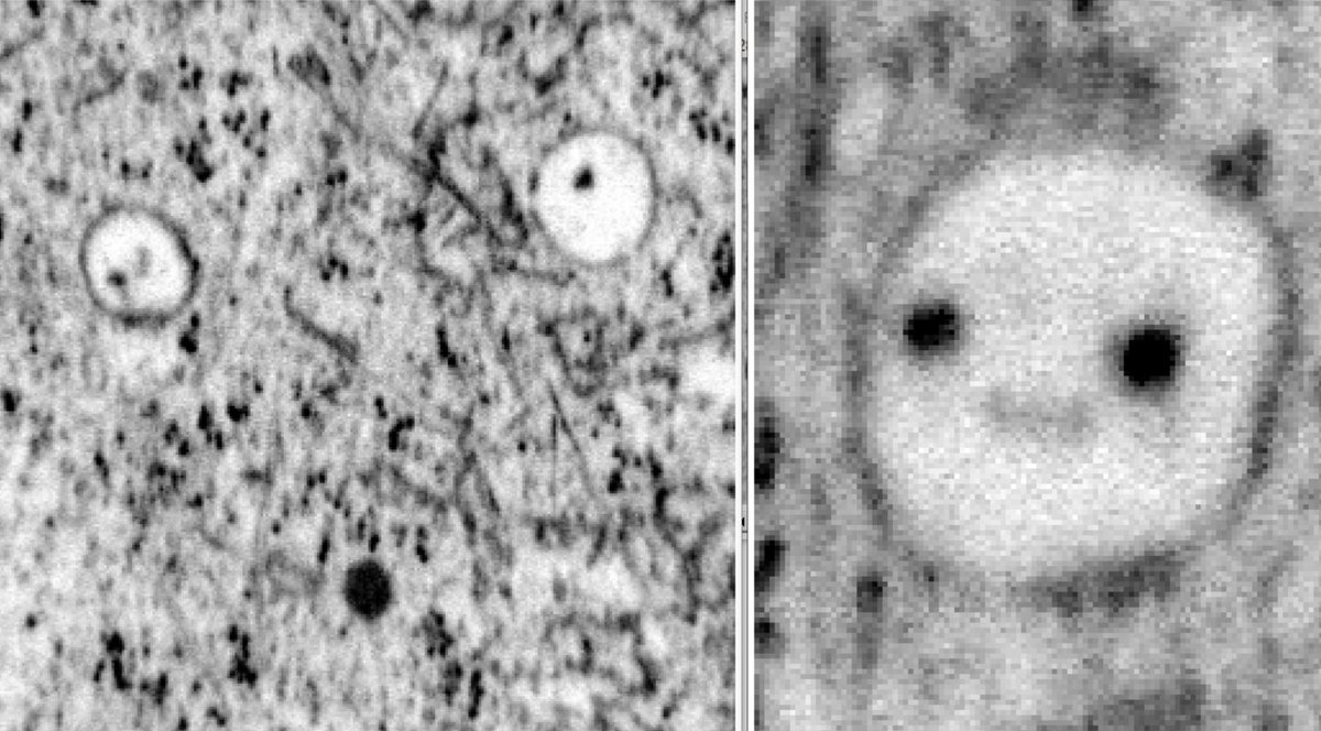
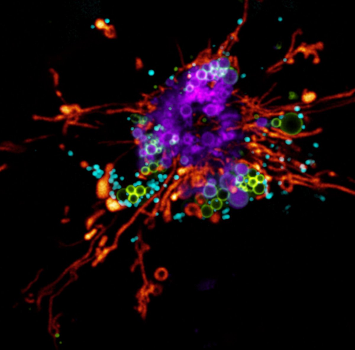
This cell has lovely bubbles and dots to go with its wormy tubes. I think I will do an image analysis on it.

This cell has lovely bubbles and dots to go with its wormy tubes. I think I will do an image analysis on it.
account_circle
Nature Reviews Molecular Cell Biology(@NatRevMCB) 's Twitter Profile Photo

✨ICYMI: New on the role of actin in mitochondrial biology, e.g., during mitochondrial fission, dysfunction and , or motility.

➡️'The multiple links between actin and mitochondria' by Tak Shun Fung, Rajarshi chakrabarti and Higgs Lab.
go.nature.com/3Xj0sMg
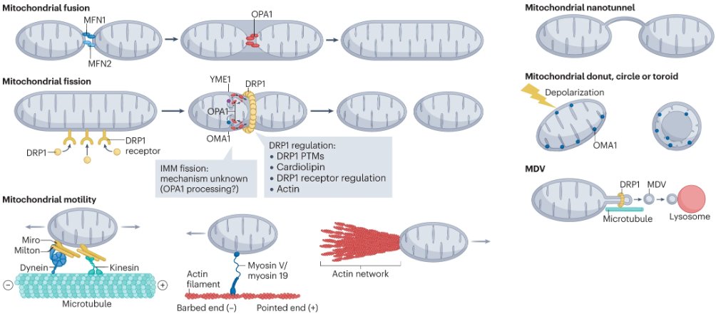
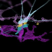
✨ICYMI: New #NRMCBReview on the role of actin in mitochondrial biology, e.g., during mitochondrial fission, dysfunction and #mitophagy, or motility. ➡️'The multiple links between actin and mitochondria' by @TakShunFung, @raj_mito and @hhiggslab. go.nature.com/3Xj0sMg
account_circle
Kowalczyk Lab(@CellJxn) 's Twitter Profile Photo

The desmosome, normally toiling away in obscurity, finds itself on the cover of Nature Cell Biology by way the ER! We are excited to see our work in press along with a News and Views commentary by Harmon and Gottardi Lab nature.com/articles/s4155…
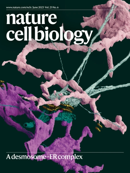
The desmosome, normally toiling away in obscurity, finds itself on the cover of @NatureCellBio by way the ER! We are excited to see our work in press along with a News and Views commentary by Harmon and @GottardiLab nature.com/articles/s4155…
account_circle
Navaneetha Bharathan, PhD(@genehackrman) 's Twitter Profile Photo

Super thrilled to share my first postdoc paper in Kowalczyk Lab lab! And I'd be lying if I said I wasn't just as thrilled about getting the cover! I'd like to thank our excellent team: Will Giang 🔬 江威廉 Stahley Lab AIC at Janelia COSEM Coryn Hoffman, Wayne Vogl... (see🧵below:)

account_circle
Till Stephan(@stephan_till) 's Twitter Profile Photo

The actin cytoskeleton of a human cancer cell imaged by super-resolution microscopy. Actin was labeled using phalloidin and imaged with a commercial STED microscope.

The actin cytoskeleton of a human cancer cell imaged by super-resolution microscopy. Actin was labeled using phalloidin and imaged with a commercial STED microscope. #STED #Superresolution #FluorescenceFriday
account_circle
Andy Moore(@aaandmoore) 's Twitter Profile Photo

Full window is 790 x 790 µm and the movie is 16 hours long at 2 min per frame. Raw movie was 280gb, this is a 6px downsample + added compression on upload.

vimeo.com/834808566?shar…

account_circle