Urmila Anandh(@AnandhUrmila) 's Twitter Profileg
Urmila Anandh

@AnandhUrmila

Nephrologist . Interested in places and people.

ID:914421804940222465

calendar_today01-10-2017 09:28:42

21,2K Tweets

3,4K Followers

666 Following

Follow People
Sanjeev Sethi(@SethiRenalPath) 's Twitter Profile Photo

It was a hard sell.
It is now accepted that the likely etiology of DDD & C3GN in older patients is a monoclonal gammopathy. And targeted treatment is treatment of choice.

But back in 2008-2009 no one would buy it. This is the very first report.

doi.org/10.1053/j.ajkd…

account_circle
Nephrology Journal Club(@NephJC) 's Twitter Profile Photo

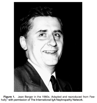
🤔 Do you remember Jean Berger from our last ? He and electron 🔬 expert, Pierre Galle, published 📃 1st description of DDD in 1962.

Read more about C3 glomerulopathy here: sciencedirect.com/science/articl…

🤔 Do you remember Jean Berger from our last #NephJC? He and electron 🔬 expert, Pierre Galle, published 📃 1st description of DDD in 1962. Read more about C3 glomerulopathy here: sciencedirect.com/science/articl…
account_circle
Nephrology Journal Club(@NephJC) 's Twitter Profile Photo

Berger and Galle described the appearance as dark, ribbon-like, electron-dense material in the central layer (lamina densa) of the glomerular basement membrane (GBM).

Berger and Galle described the appearance as dark, ribbon-like, electron-dense material in the central layer (lamina densa) of the glomerular basement membrane (GBM). #NephJC
account_circle
Nephrology Journal Club(@NephJC) 's Twitter Profile Photo

Urmila Anandh As per this seminal paper by Thomas et al., doi.org/10.1016/j.semn…, deposits can be seen in mesangium and Bownmann's capsule as well in DDD!
May be pathology experts may help out here!

account_circle
Nephrology Journal Club(@NephJC) 's Twitter Profile Photo

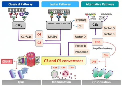
T0f: C3G is mainly caused by dysregulation in the complement cascade, this can be from genetic 🧬 and/or acquired 🧪 factors.
More than 90% of C3G cases are caused by dysregulation in the alternative complement cascade, while 10% are in the classical/lectin pathways.

T0f: C3G is mainly caused by dysregulation in the complement cascade, this can be from genetic 🧬 and/or acquired 🧪 factors. More than 90% of C3G cases are caused by dysregulation in the alternative complement cascade, while 10% are in the classical/lectin pathways. #NephJC
account_circle
Nephrology Journal Club(@NephJC) 's Twitter Profile Photo

T1e: Proteins & peptides’ intensity based absolute quantification (iBAQ) values were compared between the two groups to calculate fold-change and P values.

account_circle
Nephrology Journal Club(@NephJC) 's Twitter Profile Photo

T1f: Various other analyses performed included - immunohistochemical (IHC) staining for ApoE, and IF confocal staining for co-localization analysis.

T1f: Various other analyses performed included - immunohistochemical (IHC) staining for ApoE, and IF confocal staining for co-localization analysis. #NephJC
account_circle
Nephrology Journal Club(@NephJC) 's Twitter Profile Photo

T1g: In traditional IF, antibodies labeled with fluorescent dyes bind to specific target proteins within the tissue sample. Confocal microscopy🔬 is a powerful imaging technique that allows for high-resolution, three-dimensional imaging of fluorescently labeled samples.

account_circle
Nephrology Journal Club(@NephJC) 's Twitter Profile Photo

T1h: Confocal microscopy produces clear images with improved resolution compared to traditional IF 🔬. Combining the techniques enables researchers to study the localization & expression patterns of specific proteins within tissue samples with exceptional detail.

account_circle
Nephrology Journal Club(@NephJC) 's Twitter Profile Photo

T2b: ⚡️ApoE⚡️
♦️DDD 🆚 controls: ~32 fold ⬆️
♦️C3GN 🆚 controls: ~5 fold ⬆️
♦️DDD 🆚 C3GN: ~9 fold ⬆️

T2b: ⚡️ApoE⚡️ ♦️DDD 🆚 controls: ~32 fold ⬆️ ♦️C3GN 🆚 controls: ~5 fold ⬆️ ♦️DDD 🆚 C3GN: ~9 fold ⬆️ #NephJC
account_circle
Nephrology Journal Club(@NephJC) 's Twitter Profile Photo

T3b: Why do those differences in the DDD deposits exist? 🤔
❇️ Osmium dependent suggesting lipid component
❇️ Apo E major constituent on dense deposits

account_circle
Nephrology Journal Club(@NephJC) 's Twitter Profile Photo

⭐️Discussion⭐️
T3a: ApoE is a protein involved in the metabolism of fats.
It has been implicated in:
🧠 Alzheimer’s disease
🫀 CVD
👁️ Macular degeneration

In the 🫘 has been detected in fibrillary, immunotactoid GN, monoclonal Ig deposition disease (and, now in DDD!)

account_circle
Nephrology Journal Club(@NephJC) 's Twitter Profile Photo

T2c: ⚡️ApoE⚡️🆚 other diseases
♦️Total spectral counts (TSCs) were used showing 35 for DDD and 0-1.5 in other diseases

T2c: ⚡️ApoE⚡️🆚 other diseases ♦️Total spectral counts (TSCs) were used showing 35 for DDD and 0-1.5 in other diseases #NephJC
account_circle
Nephrology Journal Club(@NephJC) 's Twitter Profile Photo

T2e: Confocal microscopy 🔬 for ⚡️ApoE⚡️
✅ Purpose: confirm LCM/MS findings

♦️DDD: Bright staining along GBM, Bowman’s capsule and tubular basement membrane
♦️C3GN: Negative staining

T2e: Confocal microscopy 🔬 for ⚡️ApoE⚡️ ✅ Purpose: confirm LCM/MS findings ♦️DDD: Bright staining along GBM, Bowman’s capsule and tubular basement membrane ♦️C3GN: Negative staining #NephJC
account_circle
Nephrology Journal Club(@NephJC) 's Twitter Profile Photo

T1a: 12 controls, C3GN and DDD cases used for initial analysis.
Take a look 👀 at the specifics of the C3NG and DDD cases.
Anything that stands out?🤔

T1a: 12 controls, C3GN and DDD cases used for initial analysis. Take a look 👀 at the specifics of the C3NG and DDD cases. Anything that stands out?🤔 #NephJC
account_circle